Sophisticated, real-time imaging during minimally invasive, catheter-based procedures has opened the door on more treatment alternatives for patients who might have had few options in the past.

Dr. Elias
The continual advances in echocardiography are making it possible for more patients than ever to be treated with less invasive techniques for valve and structural heart problems.
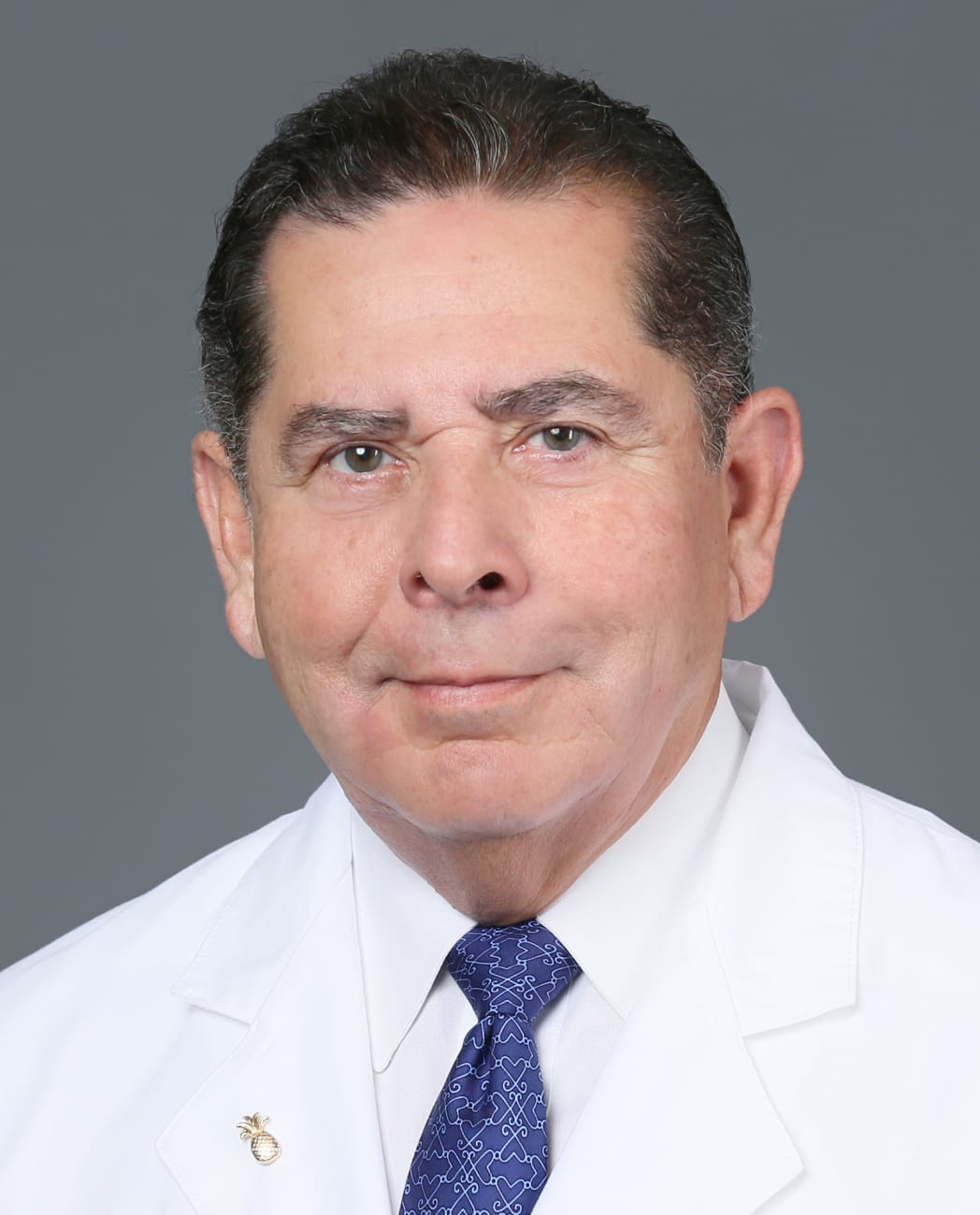
“We can accomplish so much with intraprocedural guidance — and that guidance is getting better and better,” says Elliott Elias, M.D., medical director of cardiac and structural imaging for Baptist Health Miami Cardiac & Vascular Institute.
At the forefront of these developments is the 40th Annual Echocardiography and Structural Heart Symposium. The symposium, taking place in Coral Gables on September 29-30, boasts an impressive lineup of internationally renowned faculty members from prestigious institutions nationwide which will focus on the assessment and treatment of structural heart disease, as well as explore core issues within echocardiography and advanced cardiac imaging.
Dr. Elias, symposium director, emphasizes the significance of collaborative efforts to advance the treatment of structural heart conditions. Such intimate gatherings as the Symposium foster one-on-one interactions and small group discussions, creating an environment where experts can share insights, exchange ideas, and collectively propel the field forward.
Expanding Technology
Clinical trials and partnerships with medical manufacturers have resulted in meaningful developments in treating or replacing heart valves, while spurring the next generation of devices.
“Catheter based heart treatments are consituously evolving. It is essential that we learn from each other on how to take best advantage of new cardiac imaging and device advancements for our own patients, but also for sharing these new approaches with the greater medical community so everyone can see the same benefits,” Dr. Elias says.
One example is the TRILUMINATE trial, an international study evaluating a device for patients with severe regurgitation in the tricuspid valve. The research is continuing, but has enormous potential, Dr. Elias says. “I think that it’s going to really reshape the landscape of how we treat that particular valve.”
Initially, a lot of the innovation focus was on aortic valve repair, resulting in the procedure known as TAVR, transcatheter aortic valve replacement. Within one decade of its approval by the Food & Drug Administration, TAVR has become the predominant solution for patients with aortic valve problems.
“Now we’re working our way down to the mitral and tricuspid valves, which are more complex. I think in the next five to 10 years we’ll be able to do very similar things, and hopefully with similar outcomes, to avoid surgery when appropriate for those patients,” Dr. Elias says.
It’s an exciting time to be working in this field, he adds.
“Structural heart interventions continue to evolve. As we’ve found alternatives to surgery for high-risk patients, the treatments have been so good we can now offer them to intermediate and low-risk patients for certain conditions,” Dr. Elias says. “Our goal is to minimize the procedural risk and maximize how fast patients can leave the hospital and get back to their lives.”
Noteworthy Developments

Dr. Lopez-Sanabria

Dr. Quesada
Vital developments in structural heart procedures have been undertaken at Baptist Health Miami Cardiac & Vascular Institute under the direction of Ramon Quesada, M.D., medical director of Structural Heart and Complex Percutaneous Coronary Intervention. For example, Dr. Quesada and interventional cardiologist Bernardo Lopez-Sanabria, M.D., who both will present at the Symposium, have been utilizing the new VeriSight Pro 3D intracardiac imaging catheter (ICE), the most advanced intracardiac ultrasound available.
Previous to VeriSight Pro 3D ICE, interventionalists had to rely primarily on transesophageal echocardiography (TEE) imaging to guide their procedures. TEE requires general anesthesia as an ultrasound probe is passed down the patient’s throat and into their esophagus, until it lies next to the heart. TEE usually extends preparation, procedure and recovery times — while carrying a degree of risk.
“In the past, the limitation of the intracardiac ultrasound is that it wasn’t capable of the three-dimensional imaging we rely upon for procedures,” explains Dr. Quesada. “This new device, VeriSight Pro, changed that. Like TEE, it enables 3D imaging, but without the need for general anesthesia.That’s very, very important for the patient and for efficiency. And that’s very revolutionary for our desire to get patients back to their normal lives as fast as possible.
Additional advances include ever-improving 3D scans, fusion imaging software that overlays echocardiogram and X-ray scans in real time, and MRIs with artificial intelligence.
“The technology is only going getting better,” Dr. Quesada says.
About the Symposium
As technology continues to evolve, patients can expect even better outcomes and a higher quality of life with minimally invasive treatment options becoming more widely available.
The advancements in echocardiography presented at the 40th Annual Echocardiography and Structural Heart Symposium hold great promise for transforming the treatment landscape for patients with heart valve and structural heart problems. As medical professionals come together to share knowledge and experiences, the future of echocardiography looks bright, with increased accessibility, improved imaging technologies, and innovative procedures that offer safer and more effective treatment options for patients in need.
For a schedule and information on the 40th Annual Echocardiography and Structural Heart Symposium, click here.

