When to Refer, When to Act: Critical Decisions in Aortic Disease Management Tuesday, May 26, 12:30-1:30PM ET
Format:
Latest guidelines in aortic disease
Case-based review and discussion
Live Q&A
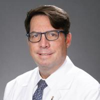
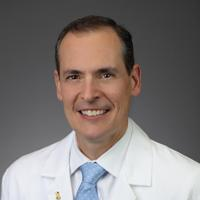
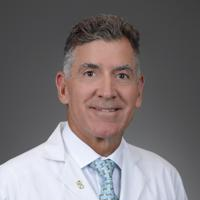
Physicians:
Dr. Ashok Kumar C J
Dr. Bradley Taylor
Dr. Brian Schiro
Dr. Ignacio Rua
Good afternoon, everyone, um, and welcome to today's, uh, webinar. Uh, the topic for today is, uh, when to Act, Venter for Critical Decisions in, uh, aortic Disease Management. Um, my name is Doctor Ashok Kumar CJ. I'm the assistant director for Aortic Disease Center of Baptist Health. And, uh, I'm moderating this today's session on behalf of the Baptist Health Aortic Disease Center. And, uh, thank you all for joining us. Um, we are excited to have Uh, physicians, clinicians, and healthcare professionals, uh, from across the region, um, with us today for what I believe will be, uh, a, a highly practical and clinically, uh, relevant discussion. Um, aortic disease remains, uh, uh, of, uh, one of the most complex and, uh, uh, time-sensitive conditions in cardiovascular care, and, uh, our outcomes often depend not only on timely intervention, but Uh, when to refer, when to intervene, and, uh, through multidisciplinary care. I think this webinar will focus on all those, uh, aspects of the, uh, multidisciplinary care, uh, the things. Um. We're honored to be, uh, uh, our agenda is to, uh, present the latest guidelines on aortic disease briefly, and, uh, we'll have a, a case-based review and, uh, our discussions of like three unique cases, what our, uh, multidisciplinary team has put up with, and, uh, the last 1015 minutes, we'll discuss about, uh, the Q&A's from the audience and we can go from there. Um, uh, we honor to, uh, be joined by an outstanding panel of experts, uh, Doctor Bradley Taylor. Uh, he's the Chief Medical executive at the Christina Lynn Medical Institute of Baptist Health, Doctor Ignacio Arua, the chief of vascular Surgery there at Baptist Health, and Doctor Brian Schiro as our interventional radiologist and endovascular, uh, um, um. A surgeon at Badress Health, um. I think, I think following the discussion. We will focus on the three cases. Uh, we'll start with the, uh, uh, the guidelines first and go from there. We hope, uh, today's session, uh, is gonna provide practical insights for, uh, people who are like, uh, in clinical practice and also those who can apply these things to refer patients, uh, to our disease-specific centers here. Uh, thank you again for being here with us today and, uh, let's get it started. So to begin, I think the global burden of aortic disease like uh there's an increased uh worldwide rising incidence of aortic disease as we speak. And the more and more it is seen on younger patients with more complex diseases and things, and there's increased redo operations as we speak about, and I think the delay seems to be or remains to be the biggest uh cause of mortality and delay in referral, delay in intervention, and delay in offering multidisciplinary care is what I believe. Uh, I mean, what do you guys think? I think you're right on all those factors, CJ. I think that, uh, definitely we're seeing more and more complex cases that are happening in, in patients, and as you pointed out, younger patients are, are getting these as well. Upfront referral is particularly important to be sure that we get these patients into a program so that, uh, so that they're cared for expeditiously. Without, without a doubt, we joke around a lot that All the easiest cases have been done. All we're getting is all these complicated cases, so it is very, very important to get a heads up on any patient as soon as we can rather than just emergently right at the end. Yeah, absolutely. Uh, you know, when I look at this slide, you know, it makes me really think that, you know, when we talk about the aorta, you have to think of the aorta and continuity from, uh, I, I, for me, it begins at the left ventricular outlook flow track just below the valve and all the way down to the femorals. So that's what we're really interested in, in, in managing and taking care of. And if you ever have a question about referring, I, I think you refer. Yeah, we want to co-manage these patients with, with the people that are primarily taking care of these patients. The field is now such that it's just amazing just to think about the aorta as an organ, correct? In the next slide, actually, yeah, that, that's actually part of the guidelines. Yeah, I think let's go for the next one here. Uh, I think, I think that 2025 is where all of our societies came up with this, uh, idea of calling aorta as an independent organ, and I think it's rightfully so. It's high time. I think it's, it was, it is a long time coming and it's finally there. I think, uh, uh, it truly requires, uh, a multi-disciplinary aortic team, uh, at Centers of Excellence or aortic Centers for lifelong management of patients and things. I think aortic disease is a, a disease of a lifetime. It's not like you're one and done and then you forget about it. Um, uh, as you see, the, it's a class one recommendation to be called as an aortic organ as for our guidelines right now, and, uh, it's also, uh, a class one indication for, uh, the referrals to multidisciplinary care and disease-specific centers for things. Uh, as in Baptist, like we have our Uh, great aortic team here, a co-radiating consists of cardiac surgeons, vascular surgeons, and interventional radiologists, and cardiologists are people who are primarily taking care of these patients. But the most important thing is that the extended multidisciplinary team, which includes, uh, cardiac anesthesiologists, perfusionists, uh, critical care specialists, geneticists, and neurologists, pulmonologists, renal specialists, specialized nurses, surveillance workers, and also a nurse practitioner who will take care of our surveillance team. I think that is what is unique about uh disease center uh uh uh like Baptist, as we have all these things under one roof. Definitely a Baptist heart and vascular care. I mean, you're not gonna find a more comprehensive group of people that are involved in caring for these patients. I think that that incredible long list of uh extended multidisciplinary team members that you have. Along with the core aortic team, it's gonna be hard to find that anywhere else in South Florida for sure. Yeah, well, it, it feeds off that whole concept of collective intelligence. So if you have a complex problem or any problem, if you get people from different disciplines and backgrounds, uh, those problems get solved in a much more expedient, and, uh, the complexity of the problem gets simplified. So when I think of multidisciplinary and what we have here at Baptist, it's really a, it's a team of, of collective intelligence. I think as, as we talk about uh referrals and planning and, and, and imaging, I think that's what we have. We have like high resolution CT scans, three-dimensional reconstructions, virtual valve sizings, AI except put a risk prediction, and digital rehearsal. We are like, like we talk about these things and uh uh uh we talk about our aortic meetings are coming, we do uh two multidisciplinary meetings, uh, uh, every few weeks to discuss and uh all these complex patients and uh as a Team, we all come together and reconstructing these things, coming with a Plan A, Plan B, Plan C, and we have everything ready for them, and I think that makes a significant impact on the successful outcomes of our patients. Would, would you say that patients are, uh, when they're referred to the Center of aortic Disease or to any of you independently, You then share that with the group, would you say that's 100% of the cases? Yes, yeah, yeah, no doubt, no doubt. I think also on the, the list of, uh, multidisciplinary care is the world renowned, uh, experts that we have in uh cardiovascular imaging. Um, uh, the images that you see here are, are, uh, very high resolution images that, uh, take a lot of work with expert technologists that can help do some of the reconstructions on this. And the uh physicians involved with, uh, uh, expertly reading these cases as well. Yeah, I can't, I can't, you know, agree more on that, on, on what Brian is saying because sometimes Ryan or like Tina or any of our international people call, uh, uh, call me or our colleagues from, uh, from the reading labs like saying, hey, did you see this? Patient yet, even before we get to know about this patient because they get called by the ED and they're looking at the CT scans and then they call the aortic team right away, and then we have a 1 to 1 discussion right away on the phone, and that makes uh the, the care coordination is very quick and there's no delay. I think, I think that, that significantly plays a part in acute aortic syndromes exactly like we speak uh in terms of uh uh type A dissections and uh type B aortic dissections too. I think, uh, um, the indications currently remains the same. Uh, we rushed to the ER every, every minute counts in type A dissections, and, uh, as for the type B dissections, there are more data coming in on when to intervene. Immediately when compared to optimal medical treatment on these patients, so like, what do you guys think on, uh, on, I mean, the, the type B, uh, dissection that is from the left subclavian and, and beyond, you know, require. You know, a lot of clinical input from many, many people, you know, many, uh, things are, uh, uh, in play as far as, you know, uh, perfusion to the viscera, profusion to the extremities. So that, that's the time period I think where it's great to have so many people involved because let's say there is an ischemic problem, well, We may need the aortic root fixed ASAP. We may need to stent ASAP and in other patients, we wait. I mean, it's just a lot of clinical decision making, um, and uh. Kind of, that's the. Uh, art of this whole, uh, uh, I think in a true type A dissection with malrofusion, I think that is something a different entity as a different beast. Uh, the only time I probably stage is when you have a mecentric malprofusion. I've got ischemia. At that point in time, I would like to rather stand it first and wait for lactase to normalize and then go and attack the uh ascending. What about you, Brett? Yeah, no, that's been a long standing. Practice, uh, that, uh, came about probably about a decade ago, you know, the, um, Obviously, if you can take any acute emergency and turn it into a more of an elective, it decreases the uh the risk to the patient as a whole. Open surgery still is the gold standard for an acute type aortic dissection. Um, there is an expanding role for stent grafts in the ace and aorta. We've learned a lot in cardiac surgery from our management of type B's, which are now primarily either medic. Primarily either managed medically, that's a mouthful, or uh with endovascular grafts, which have really kind of uh uh revolutionized the care of, of type B aortic dissections and quite frankly, aortic pathology. Sounds good, sounds good. I think next one is. I think like we talked about, I think this chart shows us exactly what uh the management of type periodic dissections um when we opt for uh optimum medical treatment versus TW in the same admission or within 3 months. I think uh we have a select, I think that our societies have come up with great guidelines, uh, to, to for all of us to, to look into and then. Uh, uh, justify with evidence-based, uh, data to when to offer, uh, TA the same setting when compared to optim medical treatment. I think, I think I can honestly say that at Baptist at all, as our aortic team are like 100% evidence-based, and we, we comply with all the guidelines and sometimes, um, we challenge the guidelines. That's very important. Every patient is different to a certain extent. Um, classically, I think, uh, this, this chart probably shows us the, uh, the current guidelines for, uh, referrals for, uh, intervention for ascending aortic aneurysms, both sporadic and bicuspid. For us as an aortic center, uh, we go anything more than 5 centimeters. Uh, we don't wait for a classical 5.5, um, but 5 centimeters, but that being said, every patient has to be tailored, customized, and discussed among multiple. It's not like if I'm seeing the patient. It's not like I'm the judge, the jury, and the executioner for that patient. I don't want to. I tell the same patients, uh, all patients all maybe, I do the exact same thing, what I would do for myself, my family, and my friends. That means I'm gonna discuss with my expert colleagues and say, hey, is this the time to intervene and is the right thing to do for these patients? Uh, that being said, I think, and so many of our patients we see are like have high risk factors, so we tend to intervene at a, at a, at a much lower threshold. Um, how about you, Brad? Yeah, so certainly if somebody has a connective tissue disorder, uh, we'll operate on those patients at a smaller aortic size, generally around 4.5, um, and in young patients with specific congenital issues maybe at 4 centimeters. In general though, well, uh, uh, we refer patients for surgery at around 5 centimeters. Now certainly if someone's at the extreme of age or extremes of disease where it puts the risk of the operation at a higher, we may let that, that aorta grow a little bit more. But again, it's all based on what's, what's in the best interest of the patient. There is a group of patients though that's going to the operating room that has about a 4.5 centimeter aorta. That for instance, is going in for bypass surgery or some concomitant uh cardiac surgery, we'll take care of that aorta at that time because we know that in general for most people, once the aorta gets to be about 4.5 centimeters in size, it grows about 1 millimeter to 1 millimeter.2 a year and so within 5 years, they may be faced with another operation and it's better to take care of it at that time than to wait. You know what I find interesting also from the guidelines is the, the fact that there really are almost two different approaches to when a patient should be treated. So, interestingly, if you look at this, uh, for a 5 centimeter aneurysm, those patients are are sent for treatment and patients in, excuse me, in institutions that have a multidisciplinary aortic team, which is exactly what, what we're proposing here. What are your thoughts about uh the benefits of that versus when they're saying 5.5 centimeters in, in a standard uh um facility or standard hospital system? You know, it, it's interesting that it, it, it. It references both. In around 2012, the guidelines was 5 centimeters for the ascending aorta. That changed around 2012. They changed it to 55, and what people found is that People weren't surviving as long, and so for patients that's the places that see a lot of aortic disease, they've gone back to 5 centimeters. And I think what we're going to see is that these guidelines are going to shift down to 5 centimeters. I, I can't speak. I wasn't part of the writing committee, but if you think about it, if you're a patient, You want to get taken care of earlier and you wanna be in a place where somebody does. If you look at volumes of centers that do, and that magic number is 20 aortas a year, it doesn't matter if it's descending, ascending, 20 aortas a year, there's a survival benefit. If you go to a center that does more than 20 ascendings a year or 20 descendings a year, your likelihood of survival is greater than if you go to a place that's less. That does less than 20. Yeah, I think that again highlights the multidisciplinary care that we have at uh Baptist Heart and vascular care. Well, the next slides actually shows exactly the same thing, probably the one next to that. This is probably your back cusp, but probably the next slide will. I think this, this, uh, chart was more interesting for me because as Brad O'Reilly said, uh, I think going intervening on 4.5 centimeters is right now a class twoA indication, and we do that all the time. And I think the most important thing for me is, uh, when I'm discussing with the patients is the bottom half of the thing that shows the risk factors. It's like, I think the length of ascending aorta is, uh, more than 11 centimeters from the, the root to the origin of the right anomin artery is a significant. Uh, a variable, that, that, uh, society is looking into more and majority of the aortic centers across the world are looking into and ours is definitely one of those things. So I look into that thing aspect definitely if the patient is borderline but has these significant, uh, characters of risk factors like. The patient is shorter, like less than 1.69 m, or like patient has a significant family history or a systemic or if the pressures are like always running in the dark, my pressure always runs in 200, yes, like, yeah, I'm gonna intervene on you, even if it is like 4.8 centimeters for the same reason. Yeah. CJ Chad GPT won't tell you that all that. Put all that together that I can tell. That's why it takes the personal touch. Yeah, I think that there's a lot of. Art and science, I'm glad you brought that up because, uh, in, in this era of AI and chat GPT and, and, and, and the knowledge is there everywhere, uh, I see a lot more patients. I would like to your insights and thoughts on things. A lot of patients actually come and tell me, OK, I need to be intervened right now because Chad GBD said so. I think, I think this, this chart actually, uh, is a very good one to discuss this because I think, uh, there, there's two. Two hinge points of inflection in terms of, uh, aortic length. One was like more than 11 centimeters and another was more than 12.5 centimeters. As the chart on the right side looks at it, the, the, the, the chance of the probability of acute aortic events significantly goes up, as you can see when it is more than 11.5 centimeters and, and then the second hinge point is more than 12.5 centimeters. I think, uh, we, we need that, that is why the true, uh, team of, uh, uh. Uh, radiology, and diagnostic radio interventional radiologists telling us the lens and discussing with our aortic team meetings, all, all, all collaborate to come and do the right thing for the patient. Yeah, think of it this way, you know, we've always thought about dilation of the aorta, the, the law of Laplace that the tension increases as it expands, right? The wall thins out. Well, the same is true if you elongate that same tissue, right? It's being stretched thin. It'd be really nice and maybe Brian can speak to this is like, are we able, will we ever be able to predict based on the thickness of the wall of the aorta, particularly the media? That's, that would be something great to look at and that's where we could actually employ some of the AI tools to, to answer some of the questions that we, we don't even know to ask yet, right? So that in particular would be something interesting to look at. We don't have any specific data on it as of now, but, uh. More to come hopefully. Yeah, I think that we're applying that thing in the aortic valves. I'm glad you brought that up because uh looking at photon counting CT scans to look at the thickness of the valves and predicting the things, that'll be a great research topic for us to go into things. That's the thing, yeah, I just want us to move a little bit, uh, into the, uh, cases. So really quickly, the descending aorta and the thoracic abdominal aorta are very problematic because these are the branch points that, you know, the, the branch, the, the, Um, celiac and superior mesenteric that, uh, perfuse the liver and the viscera, and then the two renals. So, these are, uh, blood vessels that need a lot of, uh, attention to getting things done fast and in an expeditious way to protect them. Um, so that's the main problem with, with these, but as, As far as the guidelines go, um, it's, it's pretty much, uh, uh, 55 millimeters. We're still with those, but again, it's tailored to the overall patient, the risk factors that the particular patients have. But as far as, uh, you know, referring the patients, you know, 4.55, you wanna make sure you're in a place where there, there's the ability to, to take care of the patient. Great. Same goes for your abdominal and infrarenal abdominal and infrarenal, same sort of, uh, these are kind of a busy slides, but the main thing is just refer the patient early so they come to the attention to, you know, a place where everything can be handled. I think we were talking about earlier like it was like a threshold of more than 3, 3.5 centimeters. And a bowel should be there for surveillance. those patients definitely should be in a surveillance, uh, to watch for aneurysm growth. One thing that we look for in particular is growth of that aneurysm of, uh, uh, 10 millimeters per year. If we see something, uh, that rapid, those patients need to be followed more closely or even offered treatment, uh, based on that, that, uh, rapid growth, yeah. I think this is a great slide. I've, I, I don't know because it's like we discussed about the, uh, aortic disease being a disease of a lifetime, and not, not just being treating or something, what to do before for some patients who are on surveillance, like what should they do, what should, what cannot, what, what can they can do. Um, I always tell them like, hey, if you have a 4 4.5 centimeter like aneurysm, he's sending aorta like and, and, uh, uh, he, he, he's one of my patients was like a skydiver. And I said, OK, thanks. So I tell him like, you know, I'm gonna fix you first because you're not gonna do skydiving. And then like, and, and, and even before we fix them, he said like in the two weeks before he was skydiving. I said you cannot control those people. Uh, and then we tell them, OK, they're, they're, don't avoid contact sports. Do not control your blood pressures, uh, or control, impulse control is very important. Do not do any heavy lifting is what we suggested before the surgeries. And the next question they ask is like, what if after the surgery, can I do it after the surgery? Well, I tell them it depends upon the, the rest of your theory dimensions and things. Yes, you can go back to your normal lifestyle, but if you have untreated connective tissue disorders, you definitely have to change your lifestyle based on. What's your recommendations? This, this is a great list because it's really, you know, these are the questions whenever the patients come into the office to see us. These are the questions that they ask, right? And what we have to encourage and what's number, uh, number 1 on the list here, it's a class 1, C recommendation is that they should continue to exercise and continue with sports because they need to keep their cardiovascular health up so that if at some point they do need a repair, they can tolerate it and recover well, yeah. It's what we call as a prehab. That's right, that's right. That's right. Stay strong. I, I advise patients that they should still lift weights and that they should exercise and exercise, you know, to, to stay in shape. It's really that Valsalva maneuver where they're bearing down to do that, that extra set. And most of us, once we get to a certain age, we run the risk of hurting our joints and our spine more than by lifting heavy, heavy, heavy weights, uh, or doing those extra reps, right? So, I want my patients exercising, I want them to be athletic, um, but I, I tell them not to do that, that really bearing down to, to do that final two sets that, that we did as younger people. I tell them to bring them on the weights and, and do more reps. Yeah, just maintain your tone, maintain your muscle, uh, very important. Uh, I think, uh, just rushing through our, uh, to the slides to get to our case-based discussions, this is, uh, uh, the personal strategy what we offer at the aortic Disease Center at Baptist Health, and heart and vascular care, uh, like, like Doctor Taylor was saying, it's from the aortic root all the way to the femorals. Uh, these are aortic root strategies, uh, as you can see, as a class one indication for these things. I think this picture is almost like I can see a layer. Of blood going across the thin membrane. We were talking about thickness of the aortic wall. I think there was just a small antima there. I can, I can literally see the blood flow from the aortic wall going across and it's pretty scary, pretty scary, and these are the patients who are walking the streets. Without doing anything, without knowing anything, and there's a ticking time bomb when we tell them there's a ticking time bomb. This is one of the ticking time bombs there and no heavy weights on this one. No, no, no heavy weight for sure. Again, there's another valve sparing a root replacement for a tri-leaflet valve, and then the next one is a bicuspid valve. Uh, we, we try to, uh, uh, spare the valves, uh, at, at. At every, every aspect we can either in tricuspid, now, especially on younger patients, uh, um, for, for the durability and things so that they're not on any blood thinners or they're not on any biprosthetic valves, I think that's the thing. And even in, even in acute aortic dissections with dissections into the root, if it's a young patient less than 60, 65 years of age, we're gonna spare that valve. Correct. Um, another thing of, uh, ascendingotic proximal arch strategies, uh, as a class one recommendation for, uh, uh, all these surgeries that we do hemi arch and open arches, uh, it's, it's a pretty, uh, decent technique, uh, what we have here. And this is another aortic arch technique like a zone 2, arch replacement, uh, with a frozen elephant trunk. This is like a kind of a, uh, um, uh, be safer, uh, from one of our aortic colleagues at the Cleveland Clinic. Uh, I think, um, it, it's getting back. I think right now they're, they're. There are more, um, what do you call those um conventional devices coming up in front of us that can do it uh on the same pattern. Yeah, industry has certainly provided aortic surgeons in the last decade with unbelievable tools, right, particularly for us to manage, um, the, the arch and the descending aorta at their first time operation. And being able to extend and you know the hemi arch, I think is going to become a a more of a historic operation that we relied heavily on. And now the ability to take care of somebody's arch and, and set their descending aorta for our colleagues is, uh, in the last 5 years has certainly become much more frequent. I, I think on the same note, it would be a word of caution is I think these things should be done only in space. Uh, a very highly specialized aortic disease centers, I think, like ours, and when we have expertise to taking care of all these things I think because if, if things go south, we have the we have the resources and the team to take care of all these things. So I think this is what we offer at our uh high volume leading edge uh urtic center at Baptist open surgeries and uh do a lot of minimally invasive uric surgeries including uh uh endovascular, uh, TBEs and uh totalological replacements and TAs and, uh, endobenthos and things. um, uh, I think without further ado, let's go back to our case discussions next, um, and go for the this is just a, a, a slight, it's kind of a Uh, shows you the, the ages. This goes from, you know, a teenager to the left to one of the oldest living Marfan's patients on the right. So that you see on the image on the left, there's a contained rupture. This was a 14 year old kid. I remember this patient, yes. Yeah, it's like, uh, earlier this year. Complaining of severe back pain and there's really nothing left to do but take him emergently to surgery. That's the middle slide where we replace it all. But lo and behold, uh, a, a, a geneticist evaluated and he had errlos-Danlos, which is a connective tissue disease. And then the slide on the right is, uh, a Marfan's patient had his aortic root replaced. I follow him for an abdominal aortic aneurysm, palpateal aneurysm, but, He had a carotid artery aneurysm that about 3 years ago, uh, ended up having to resect it and Brad and you guys had mentioned how thin it was translucent. That was, I mean, really, really scary. You're asking how old was this patient? I think he's in his late 80s now. I saw him in the office maybe about 2 weeks ago. Great. He's an attorney who still practices. Awesome, and he's just, uh, very, very, very smart and takes care of himself and enjoy enjoying his life. you want to just go through the keystones, we'll get in, uh, get into some of the cases, uh, that we've done recently. So this is a 78-year-old female, uh, that had an aortic dissection, presented about 3 days of hemoptysis and shortness of breath. This patient's had, uh, a lot of previous aortic work, as you talked about, uh, and some other, uh, patients, but this patient came in in 1998 and had a type A aortic dissection, underwent, uh, repair and mechanical aortic valve placement for that. 2 years later, the patient had aortic dissection and aneurysm and had a stent graft placed for growth of that aneurysm up to 6 centimeters. She then comes back uh with these complaints of hemoptysis and shortness of breath. And here's some of the, the imaging that shows that previous graph that she has in place that, uh, you know, the descending thoracic aorta. And there's that uh aneurysm or pseudoaneurysm arising from the aortic arch. It's a little hard to see on, on these images that are a little off center, but that, uh, where the green arrows are pointing to there is the pseudoaneurysm just proximal to the um origin of that stent graft and just distal to the subclavian artery. So, in this particular case, from an endovascular standpoint, we were able to uh access the, the right groin. Uh, we came up and then also got access into the radial artery on the left side. We advanced the wire and catheter down that radial artery and, and, uh, then pulled the wire through and through from the radial artery all the way out through the right groin. After that, then we slide up this uh uh new stent graph that's coming up. And here's what it looks like after that stent graft is deployed. So, uh, this is a unique graph that has, uh, this pre-made fenestration or this pre-made portal that allows us to put a second stent that's going here into the subclavian artery. So now that has, uh, uh, uh, mostly sealed. Uh, that pseudoaneurysm, as we saw previously. So it looks pretty good on that final angiogram that you have there that there's no evidence of a leak, right? So you left it, you left satisfied and what's really remarkable about this in 2000, that was at the very beginning of, of, of thoracic endovascular repair, right? I mean. The grafts were only probably just a couple of years out and certainly they weren't FDA approved here in the United States, not for another 7 years. So this is a very early device that probably had a lot of rigidity to it, probably some bare metal stents that may have caused that pseudoaneurysm. Definitely required a cut down to get access to the groin at that time. Most of these now we can do percutaneously. It's amazing. That's such a great result there. My life is amazing. So this does show the importance of uh surveillance imaging in this follow-up CTA shows that there's still some contrast within the, the lumina of that pseudoaneurysm. So even though it's been excluded by the, the endograft with the, the stent in the subclavian artery, there's still some flow in that pseudoaneurysm sac. Now, as you can imagine, this does create a, a bigger problem because now, uh, accessing this uh through the blood vessels or through the aorta itself is gonna be much more difficult. So what we did in this particular case is we used uh some advanced fluoroscopy imaging techniques that includes a cone beam CT which allows us to take a CT scan of the patient while lying on the fluoroscopic table. We can take those images and fuse it over the the floral images, which is what this purplish is, uh, purplish color is. And then we can actually use guidance software in order to access that pseudoaneurysm sac. So what we're doing is uh percutaneously putting a needle from the chest wall directly into the aneurysm sac, and that's what you're seeing here on the middle image. That's great. That's great. Again, Brian, I think that not only uh reinforces for me by looking at this thing is like how much of the great outcome we're doing, but like I said, uh, uh, the multidisciplinary team approach, uh, I forgot to mention is like, you can have a team, but you also need a place where the infrastructure is there, we forgot. I think this is the infrastructure where we can do all these fantastic. In a way to, uh, technologies there like in our center, like you can superimpose a fluoroscopy and a CT scan on a patient to, to get what we want and, and, and lead the way and to treat these patients. I think that's truly, truly uh a commendable thing. Yeah, the, the tools and image guided therapy these days is really fantastic, and we're, we're fortunate again at Baptist Heart and Vascular Care to be a center where we, Uh, have the ability to test a lot of the equipment before it's even commercially correct, right. So, uh, once we're able to get into the aneurysm sac, you can see it here in fluoroscopy where we're injecting contrast through that needle. You can see right where that is, and then we treat that with, uh, putting some coils or little wire pieces into the aneurysm followed by a glue type material, uh, that, uh, that really seals off the, the remainder of the flow in that aneurysm sac. That's what the, the dark uh. Dark cloudy stuff here is in the in the center of the image. Nice, nice. How many, uh, coils do you guys throw in? Uh, we put in, uh, actually not too many because the coils these days are, are relatively large, so it takes, uh, probably about 3 coils, maybe 4, but really the, the biggest part here is putting in that liquid and bolic material that, uh, that seals off that. Is there any specific care that you guys take to make sure there's there, not a spill off or anything there? What is it, can you briefly, yeah, we watch very closely under fluoroscopy, you know, where this is live guidance so we can see what's happening, uh, the entirety of the time that we're injecting this, and, uh, uh, it gives us a, a great picture of what's happening. Here's the follow-up imaging afterwards that, uh, uh. That shows that contrast now that's sitting up in the aneurysm sac. You can see in the first image, that reconstructed image, but on this follow up. Already an early decrease in size of the aneurysm sac. We've got a CT scan here that that shows the the rest of that. Again, no flow in that aneurysm any longer. One of the good things about that, uh, material that we put in here is that it's so dense, it's difficult sometimes to see what's happening around it. But, uh, but this was a, a great outcome from that. Beautiful. And postoperatively, the patient actually did very well. No recurrent hemoptysis after the two treatments that the patient had. The patient was able to transition to full dose anticoagulation for the mechanical heart valve. Uh, the patient followed up two months later and was doing, doing extremely well. It's amazing. How do you plan to follow them up? Like, what's your surveillance method for this? So typically what we do for surveillance after we uh treat these patients is, um, we get a one month follow up typically just as a reset so that we know how things look. We then get a follow-up study at 6 months. If everything's stable there, we follow patients yearly afterwards with, uh, with repeat CT scan. Again, that surveillance is particularly important because we want to be sure that there aren't any complications that's occurring after the aneurysm repair. That's particularly important with patients that have, uh, endograft repair. Any strategies on anticoagulation on these patients? Uh, uh, just like this patient, the patients can go back on anticoagulation afterwards. We don't typically put patients on anticoagulation if, if they were not on it, uh, preoperatively. Uh, it is important for cardiovascular risk factors, of course, for patients to be on the appropriate medications, which usually include, includes a low dose aspirin, and then a lot of patients go on, uh, rivaroxaban 2.5 mg, uh, twice daily as well. Uh, some other patients are managed with Plavix if they have, uh, uh, clopidogrel if they have other, uh, other reasons to be on it, but for the most part we don't put patients on anticoagulation unless, uh, required for other reasons. So the same strategy for your practice, Brett, that's correct, yeah. Let's go on for the second case study there. So this next case, uh, to talk a little bit more about the abdominal, uh, aorta. This is a 77-year-old male that actually had a surgical repair of an infrarenal abdominal aortic aneurysm 15 years ago. And now I was talking about surveillance and I was saying that uh in general, it is important for us to follow up patients that are post stent graft, but even patients that have open repair uh do at times have recurrence of uh aneurysmal disease. So this patient did have a previous repair and presented acutely to the hospital with flank pain. This is the initial scan when the patient came into the hospital. And this is a device that's available on the from the FDA now. This is a multi-branch device. It's called the thoraco abdominal branch endoprosthesis. What it has is uh uh portals that we can place these stents through in order to maintain blood flow into the visceral vessels. That first CT scan was a surveillance scan that the patient had, and then he came back with this scan. Uh, this is the initial one when he presented to the hospital, and it is certainly different in appearance and the fact that the aneurysm is bigger in size. There's also what the green arrow was pointing to here is some stranding around the side of the aneurysm. This makes us concerned that this may be an impending rupture. In addition to that, this patient had acute occlusion of the left renal artery, so you're gonna see there's no enhancement in that uh left kidney, whereas the right kidney still has a good perfusion. So these are the things that we need to think about as we're going in to potentially treat this patient, particularly with that Tambi device. And again, here's the um uh stranding along the walls of the aorta, really certainly making us concerned about potential rupture. So we wanted to treat this patient expeditiously. This requires, uh, uh, really two very important accesses. Uh, this first access is in the right groin. Uh, on the far right, you're gonna see some, uh, uh, um, surgical instruments there because we also got access into the subclavian artery via a cutdown. There's the access into the groin. And then we have wires going through the blood vessels. Again, the importance of a multidisciplinary team approach, you can see the large number of people that are involved in this particular case, and the reason for that is because it's extremely complex and requires multiple specialties and multiple different operators in order to perform the procedure. They always say it takes a village to, to, to do these surgeries and procedures, and then this is definitely one of those. All right, so, uh, once we have, uh, um, access, here we are, uh, snaring the wire. So again, we have access now all the way from the subclavian artery all the way through the right common femoral artery. And you can see the arrows there pointing to the, the wire that's been through and through. Once we have that wire through, then we load in multiple other wires for a total of about 5 wires that are sticking out of the patient here, and that's why we have uh that little white device there in the center to help hold some of these wires, uh, and try to keep everything straight. Requires a lot of labeling and a lot of uh a lot of patience definitely to get to that point, correct. Once we have the uh uh initial device in, we partially deploy it, so then we can get into the visceral vessels. That's gonna be the uh superior mesenteric artery there, excuse me, that's gonna be the celiac artery at the top there. Uh, you're gonna see wires in the uh superior mesenteric artery as well as the right renal artery. Remember that the left renal artery was occluded here, right. That's followed by stents that then go through the portals. We deploy the stents in each one of those branches. Uh, and then we extend the, the endograft down into both limbs on the left and right side. We balloon all of the uh segments, and then after that, you can see this is what it looks like. So maintains excellent perfusion into the celiac superior mesenteric artery and right renal artery uh with uh with a great outcome here without uh any significant endo leak. And we were the, the first to perform this in South Florida, so we're pretty proud of that, uh, uh, again, showing the great work from the multidisciplinary team here of, uh, of treating these patients. Great outcome. It's a great outcome there, you know, which otherwise would have had to been repaired with open surgery, bypass, bypass all the way from, you know, you would have had to probably take down the diaphragm to get control of the proximal aorta, proximal descending aorta. It's a, it's a redo the operation, yeah, yeah, I think it's still a very morbid operation to do with these things open and like with abdominal or even some of these X and 2s and Xs and threes, uh we do like I think these novel devices that are coming in. Uh, a team approach what we're having is avoiding these morbid procedures. Yeah, I think this new technology really gives us the ability to treat a lot more patients that otherwise wouldn't, wouldn't be offered treatment. Correct, yeah, this patient, I mean this is a patient that had a previous abdominal surgery, uh, aortic repair, so this would be a redo operation, yeah, yeah. Well, I mean, like Ignacio said, like we're from the beginning of the webinar, like we are at the baptism and one of the blessings and the curse of being an aortic. Disease center, specialized centers like you just don't see the routine bread and butter stuff. You see a lot of redo surgeries, redo things like one of our patients we were discussing like we've been done like 3 or 4 times, uh, so 3rd or 4th redos and, and that's why we, we are experts in taking care of it. I think, uh, that is, that, I think the more we do it, we, the, the better we get it. The final case, sorry, go ahead. Yeah, no, your next case kind of highlights how, you know, that was a commercially available device, and the second case is going to highlight the fact that we can actually take commercially available devices and modify them based on what a specific patient needs. And that's why I like what you have highlighted here. So we'll, uh, get into that in this case. So this is a 50-year-old patient that had, uh, chest pain and abdominal pain on presentation. Had a similar episode back in 2020. This is the, the first uh evaluation of the patient in 2020 where you can see the patient has an aortic dissection as well as aneurysmal dilatation. Uh, definitely the aortic dissection is uh complex and the fact that it goes through the abdominal aorta and as we see here, this is going to continue all the way down to the, the visceral segments. Um, these are incredibly difficult to treat, as we've been talking about with, uh, with standard techniques and even some of the, uh, devices that are commercially available now have limitations that prevent us from getting everything done that we need to get done in order to effectively treat the patients. So fortunately, we have, uh, some great surgical skills that will help, uh, that we'll highlight here. So just to quickly go through a very important part of this is that the patient had, uh, in 2020 did have a treatment of that. Uh, uh, first portion of the aorta with A branched device. This is one that we talked about previously that has a stent into the subclavian artery. You can see there that the the aorta below this still is aneurysmal, and it's gonna require a second procedure after this. So, the next plan for this patient was to uh to treat the pervisceral segment. We saw that the aneurysm and dissection occurred from the distal descending thoracic aorta uh to involve the, the uh suprarenal abdominal aorta, but in order to get an appropriate seal of that, there aren't any standard devices available. Here's the, the angiogram really highlighting the fact that there's a a big aneurysm here. That is just above the the visceral segments, but also does include that as well. So Brad, this is what you were talking about as far as other options that we have for patients. When we don't have commercially available devices to treat patients, this is gonna be our, our next step, and this definitely requires lots of planning, lots of uh imaging techniques, and lots of preparation from uh uh surgical skills uh to help build these devices as well. So you can see that you've created the fenestrations that are gonna align with your visceral vessels that you could then stent into. Which is, uh, you know, we can do this, we do this type of work in the root, in the arch, and also in the thocal abdominal region of the aorta, uh, and it provides a nice, uh, alternatives. I think eventually, um, as companies and the engineers get better at making devices and we've seen a great evolution of these devices, these will then, they'll be, uh, modular, uh, component graphs that we can then, we won't have to have physician modified, but that's what. A piece of technology, it's like every 6 months there's something new that we can take advantage of. It's a great time to be doing this. It's a great time to be in the aortic, uh, uh, realm because I think finally like what I was saying, industry is taking notice of the importance of these devices for these patients. I think there's a lot of investment from industry, uh, to, to come up with some, some devices that's helpful. OK, so, uh, we'll go through this pretty quickly, but here's the deployment of, uh, uh, one piece of the endograft that we needed to place first. This is the physician modified endograft that is going in next. And, uh, our goal here again is to, uh, put wires and catheters through those fenestrations or those holes that were created surgically, and then to place stents in each of those in order to uh get an adequate seal to exclude the aneurysm. And that's what's being done here. So lots of wires and catheters, lots of technique. Lots of uh great imaging, that's all part of uh getting us here to where we need to be in order to to do this treatment for this patient. And you can see there that's a great outcome with good flow and all of the, the visceral vessels, no end of leak, uh, identify the conclusion of this procedure as well, and the follow-up CT scan really shows complete exclusion of that, uh, aneurysm and dissection, and, uh, the patient has a stent from the subclavian artery all the way down to the infrarenal abdominal aorta with these uh segments in each of the mesenteric vessels with good flow. That's, that's a beautiful image showing all those tests, uh, physician modifest things there, um, and this patient, uh, did great afterwards. Uh, we did place a, a spinal drain in this patient, you know, spinal cord ischemia is something that is important for us to always keep track of and, and, uh, uh, always keep in the forefront of our mind whenever we're treating these long segments of the aorta. Uh, we want to prevent, uh, the potential complication. OK. That's excellent. I a great case again like for things. I think, uh, uh, uh, it's, it's, it's a great presentation of these three unique cases that we have done, uh, when, in, in respect to time and we can, we can show like hundreds of these cases that we do in our aortic center. I think, uh, as we, as we do more of the AODC uh uh webinars, we'll be sharing more of the open and hybrid options in the future like in uh once or twice or 3 times a year, come up like it as a team and to showcase what we can do. I think one of the questions uh is like, um, Uh, if you have an aortic aneurysm, what can I do to decrease the risk of aortic rupture or dissection? Are there any medications or exercises I should avoid? This is the question for me. I, I can take that one. Yeah. So I think first and foremost, if you know you have a diagnosis of an aneurysm, you should. If you're not being referred from your primary care physicians, you should seek it out yourself. There's some point in time, as, as a, as an adult, you just got to take care of your own health. And so I would refer myself to an aortic center and have annual surveillance of my aorta, whether it's 4 centimeters or 4.8 centimeters. And if it reaches a size where it should be treated, you want to be in the hands of somebody that already knows you. Obviously, taking good care of yourself is the other. If you smoke, you gotta stop smoking, cause we know through lots of VA studies and studies of smokers that once you stop smoking, aneurysms tend to stop growing. And so, and the biggest cause of atherosclerotic disease and aneurysmal growth is smoking. That's one of those things. So, and then the other is blood pressure control. So I'm gonna give you three simple things. I think everybody should have in their bathroom the same place where they brush their teeth, a way to measure their blood pressure, and you should record your blood pressure, know what your blood pressure is, and if you're hypertensive or you have some evidence of it, you gotta get, you gotta seek therapy. Yeah, I agree. Anything else? No, I think, uh, those are real, real world scenarios, real-world points that you're clearly mentioned, Brad. I think is, uh, I, I do the same thing to my patients too, and then, uh, the most important thing is early referral. Early diagnosis and early intervention on these patients. Um, we, we talk about the size criteria, OK, is it like more than 5 centimeters or 5.5 centimeters, uh, does it cause an increased incidence of type A dissections? The data shows, yes, there's inflection points at like more than 5.5, 5.2 centimeters, but like you and I have seen it multiple times. Yes, have we seen a dissections on a patient with 3.8 centimeters? Yes. Have you seen it 4.2 centimeters? Yes. Have you seen it like patients walking the street without dissection at like 6.5, 7 centimeters? Yes. So I think uh the guidelines give us, uh, uh, uh, the guidelines, the, the, the spectrum of things. But to your point, the thing is like, uh, uh, constant self-awareness. And self-monitoring and if somebody is not referring you to a specialized addict center like volunteering yourself to get yourself in the line and get checked up or surveillance, it is something that awareness that we need to do, um. A couple of important things. One is, there was a study from around 2016 in Tokyo. All patients found at home that had passed away were taken for a CT scan, and 12% of those patients had an acute aortic issue that was the cause of their death. Second, if you Have an acute aortic issue. Once it occurs, 90% of patients never make it to the hospital, and of the 10% who do make it to the hospital, it depends on the degree of male profusion that they have, whether they're going to survive or not. If you're not male perfused, your survival should be somewhere between 4 and 7%. But if you're, and the majority of people have some degree of male perfusion, your mortality goes up to 18 to 20%. And, and above, and if you're severely male profuse, it's way over 40%. So you want, you want to be taken care of if you have an aneurysm or an aortic uh history in your family history electively, because your risk of mortality with an elective operation is 1 to 2%. So a big difference. Other thing question we can take is like uh uh is a comprehensive aortic disease center where we have these specialists like us taking care of, uh, these patients working together as one team. Can this change patient outcomes and aortic care? I think we, uh, the answer is yes, definitely thinks. Are there measurable benefits in survival, recovery, or decision making compared to more siloed approach? But yeah, I go to guys one by one at a time on this question. I would tell you without a doubt that is correct. I mean, practice makes perfect. Practice as a team, you, you know, that just you, we're comfortable working with each other. We're comfortable working with the, uh, the scrub techs, the nurses in the room, the perfusionists. This is all 111 big thing and also, In the postoperative care, I mean you, you, you deal with very, very talented intensive intensive care team, nurses team. I mean this is the whole spectrum, and that's the beauty of, of having a center like this. So 22 thoughts. One is, the American Heart Association's clearly published data that shows that magic number of 20 cases or more at center. those patients who aren't expected to live actually survive, and patients do better. So you want to go to a center that does at least 20 or more cases a year. And then the second, what, what separates the high-performing cardiac surgery or cardiovascular aortic centers versus those that aren't as, you know, just your run of the mill, uh, uh, centers and its ability to rescue patients. So cutting in the sewing or putting the catheters and stents in patients is actually in many respects, the more simple part. It's identifying patients before they get into trouble and their life's on the line and rescuing them before they're at risk of death. And so I think that's the difference, and that's what you were highlighting when you talk about the perioperative care, postoperative care. Yeah, it's the collective decision making. I mean, having multiple specialties involved with the care of the patient is really going to provide. The best of all worlds for the patients because they are being approached from multiple different angles as far as what's the best care for the patient, planning the the appropriate treatment for the patient, and then executing that treatment, uh, uh, having multiple specialties involved really is a a um. Is gonna improve the outcomes for the patients. The patients are gonna, uh, do better. They're gonna recover faster. Uh, obviously, the, the more, uh, data that we have, the more we're gonna see that, uh, uh, outcome. Yeah, I think on top of that, what I would like to, uh, uh improvise or not improvise. The thing is like at our aortic center at Baptist AOC Cents, what we are looking into one of the key performance indicators are like, I would like to take 31 is from the patient coming in the type A dissection in the ED. The time to diagnosis. And time to decision making and the time to intervention or cut etc. These are three key performance indicators that we as a team look into ourselves for quality data. So, I mean, even in the last few cases that anything was under 40 minutes. One of the patients was like under 40 minutes from the ED to the operating room was like that included, uh, uh, one of the patients had to go to the CT scanner and then go. We were like waiting in the ED when this patient refer came in and scooped him out into the OR and that makes it. A big significant difference on these things. Maybe we can dig one more question bigger advances, the risk stratifying question there. So it's important for everyone to know that, um, all of our patients can be evaluated because we have an STS database and you have an SVS database where we can actually impute characteristics of that patient. And then get real numbers based on comparison to hundreds of thousands of previous patients that show us what their mortality risk is, morbidity risks are, and we can have honest conversations with the patients. That's true. That's true. I think, again, I think in respect of time, uh, uh, it was a great discussion with, uh, um, all of us, uh, being here and thank you so much for all of you, uh, taking time from your busy schedule to come and, uh, uh, be part of this great, uh, aortic webinar series. I think it was one of the beginning of the many, uh, series that we have planned to do at least every few months, uh. Going forward, I think, uh, the next time we're gonna discuss more about, uh, open cases and hybrid options and endovascular options apart from the multi-trials that we are like trying to do here. And, um, is there, is there something that you guys like to say to the referring physicians uh or to the audience right now to just wrap it up and like. Well, I do want to point out that we have a referral website here. If you have any questions or if you'd like to send patients to baptisthealth.net and heart referral or you can send them to baptisthealth.net aortic care. Uh, there's lots of information on the website and, uh, uh, we look forward to, to caring for your patients. I think we have a 24/7 hotline for aortic care right now at Baptist, so all referring providers can call anytime to the 24/7 hotline and refer patients whether it's an acute, when they, they like Brad said like when in doubt, just send the patients here. We'll take care of them and our job is to take care of their patients, treat them and give them back to you. So that you can take care of them in the community and get them back to their meaningful life. Happy to answer any questions at any time. Yeah, right, right, thanks. Well, again, thank you so much again for, um, joining us. I think it's the first time, I think all 4 of us, uh, finished the, uh, webinar on uh in more than 1 hour. It's a good thing. I think, so 10 points for Gryffindor. Uh, on that, yeah, thank you so much for the Baptist, uh, group, uh, for organizing this webinar.