Pituitary Apoplexy: Lessons from Two Complex Cases
Live Webinar for Clinicians
Originally Recorded: Monday, September 15th | 3:30–4:30 PM | Virtual
Pituitary apoplexy is rare, dangerous, and demands rapid multidisciplinary action. Join Evan D. Bander, M.D., Alissa Kanaan, M.D., and Michael Fili, M.D. as they share two real-world cases — from sudden onset to diagnosis, endocrinologic stabilization, surgical intervention, and recovery.
What You’ll Learn:
How to quickly recognize the signs of pituitary apoplexy
Best practices for urgent hormone replacement before surgery
Surgical strategies for optimal recovery
Real patient outcomes that highlight teamwork across specialties
Case Highlights:
Case 1 – Post-Operative Surprise
A 62-year-old patient, hospitalized for aortic valve replacement, developed confusion and double vision in the post-operative period. Imaging revealed a hemorrhagic pituitary mass causing cranial nerve compression and pituitary hormone insufficiency. After endocrinology-guided hormonal stabilization, the patient underwent an endoscopic endonasal resection with decompression of the optic nerves. His recovery shows slow but steady improvement in cranial nerve function.
Case 2 – Known Tumor, Sudden Decline
A 76-year-old patient with atrial fibrillation on anticoagulation and a known pituitary macroadenoma presented with progressive weakness, confusion, headaches, vision loss, and ptosis. Imaging confirmed hemorrhage into the existing tumor. Following pre-operative hormone replacement, the patient underwent endoscopic endonasal resection with fat graft and lumbar drain placement. Despite a prolonged hospitalization, his follow-up demonstrated significant recovery in cognitive function, vision, and cranial nerve palsies.
Meet the Experts:
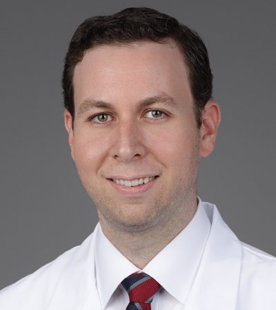
Evan D. Bander, M.D.
Don’t miss this opportunity to learn from complex cases that could transform your clinical decision-making.
Hi, everybody. Thank you for joining us. We're here to talk about pituitary apoplexy and some lessons from two complex cases that we had here at Miami Neuroscience Institute in Baptist Health. So thank you for joining us and uh we'll get started. I'm Doctor Evan Bander, I'm one of the endoscopic endonnasal surgeons here at Baptist Health, and I'm joined by Doctor Alissa Cannaan and Doctor Michael Fey, and uh we look forward to having this conversation with all of you. So as I mentioned, um Evan Bander, I'm the director of endoscopic skull-based surgery, co-director of the pituitary tumor program, and a neurosurgeon here at Miami Neuroscience Institute and Miami Cancer Institute. It's my left. Good afternoon, everyone. I'm Alyssa Canan and uh I'm an ENT trained in rhininology, allergy and skull-based surgery. I'm part of South Florida ENT Associates here in Miami. Hi, I'm Michael Fey. I'm an endocrinologist at Baptist Health and in the outpatient setting as well, and we see a number of patients in the hospital who have had pituitary surgery. So, you know, we're here to talk about pituitary apoplexy in particular, but really, uh, anytime you're dealing with the pituitary, there are a number of different pathologies that can occur within the region of the cella or uh the paracellar region, and three of the most common pathologies that we see in the cellar region are pituitary adenomas or pituitary neuroendocrine tumors, pit nets, which is what they're now called. Uh, another pathology that we do see in this region are craniopharyngiomas, which arise primarily from the infindibuum, uh, but also occur within the cella and the supercellar region, and then Rathke's cleft cysts, which are a congenital embryologic remnant, which can also occur within the cella. Now, all of these lesions can be common and need to be distinguished anytime you're dealing with a a tumor within this region. Now, pituitary adenomas or pit nets are actually quite common when they come to brain tumors. They're the second most common benign brain tumor that we treat as neurosurgeons, the most common being meningiomas, but almost 17,000 patients a year are diagnosed with pituitary adenomas. And over 90% of these are benign tumors. There are the rare few that are anaplastic or carcinomas, pituitary carcinomas, but those are very rare. We do see different types of adenomas and we'll talk about some of these, uh, including functional adenomas which are secretory tumors producing excess hormones, which include prolactinomas, growth hormone secreting tumors, which can cause acromegaly or gigantism, ACTH secreting tumors which can cause Cushing's disease. And then there are nonfunctional tumors which do not produce excess hormones and are also, um, you know, present in somewhat different ways because these grow to larger sizes often before they cause any symptoms for patients. And so to really understand pituitary pathology and how these uh how these tumors present, you really have to understand the anatomy of the region. So pituitary tumors or the pituitary glands, it's within the cellar, which is at the base of the skull, and on either side of the pituitary gland is the cavernous sinus and the internal carotid arteries. In the cavernous sinus wall run multiple cranial nerves, including the 6th cranial nerve, the abduction nerve. Uh, as well as the trigeminal nerve branches of the trigeminal nerve, and then the optic chiasm and the optic nerves run directly above uh the cella and you can see that highlighted with a red arrow uh on this MRI on the right side of the screen is the optic chiasm. So pituitary tumors as they grow, can compress these structures, and that's some uh some of the ways that these tumors can present. Now, anytime you have a pituitary tumor, then the reason that we're all here together is that pituitary tumors really require multidisciplinary care. The pituitary is known as the master gland, and endocrinology workup is really the initial first step, and we'll talk about some of that during our uh case presentations today. In addition to that, you often need input from our neuro-ophthalmology colleagues, uh, to assess the uh baseline vision in our patients through multiple techniques including Humphrey visual fields, OCT or optical or um coherence tomography to evaluate the uh the. Optic nerves and whether there's been any damage to these optic nerves, as well as neuroradiy is important in helping us interpret our imaging. And then the surgery, which is done between both myself and Dr. Kanan, uh, neurosurgeons and ENTs work together on these cases to get optimal outcomes, and we'll talk about how we work together. So treatment of pituitary tumors really can depend again on the type of tumor, non-functional adenomas or functional adenomas and depend on the size of the tumor as well as the symptoms that patients present with. And now to get to sort of the focus of our talk, which is pituitary apoplexy. So to define that really this is the bleeding or ischemia of the pituitary gland or pituitary tumor, which results in a rapid expansion of the gland and mass effect on surrounding structures. And that's what causes the clinical syndrome of pituitary apoplexy, which can include things like sudden onset headache, visual disturbance from acute uh compression of the optic chiasm or optic nerves. It can be associated with cranial neuropathies from involvement of the cavernous. Sinus and the nerves that travel within the cavernous sinus, it can cause hormone deficiencies, altered mental status, uh photophobia from uh meningeal irritation, uh, from this hemorrhage, and some bleeds, which can occur, can also be subclinical. And so we're here to talk about the management of pituitary apoplexy and we'll do that through our cases. So the first case that we're presenting, uh, is a 62 year old uh male patient who had diabetes, hypertension, and was actually within the hospital, uh, undergoing an aortic valve replacement. Pooperatively, he developed confusion, double vision, um, and on workup he underwent a CT scan, which was done by his hospital medicine team. On our exam when we first met him, he was alert oriented times 3, but he was sleepy. He was he was mildly confused, um, he was forgetful, and in addition to that, he was noted to have bi temporal hemianopsia, which is vision loss within the periphery, uh, the bilateral periphery. He had an acute cranial nerve three palsy with ptosis of the right eye, as well as an acute, uh, abducent's nerve palsy with double vision on rightward gaze, uh, but otherwise he was full strain. So, you know, when we have this patient present, uh, the first thing that I do is call Doctor Feely from endocrinology. So, a patient like this presents with, you know, sudden changes that are suggestive of apoplexy. The most important thing to do is evaluate the pituitary adrenal axis, that is cortisol production under the direction of the pituitary. The other pituitary hormones are not critical hormones. Their deficiency does not need to be treated acutely, but these patients If they become acutely adrenally insufficient, can develop shock, uh, electrolyte abnormalities, and the, the best thing to do when you see these patients is get a stat ACTH and cortisol. The other laboratories certainly can be sorted out later. His presentation was very suggestive of pituitary. Whether it was chronic or acute is is hard to tell, but some elements of it suggest that there was some crinicity, specifically the testosterone level of 8. If, if you develop sudden pituitary uh insufficiency, you have a lot of bound testosterone circulating in men, and it'll take some time for the testosterone level to get that low. In addition, the thyroid, if you look at the thyroid function studies, his TSH was in the lower ranges of normal, and thyroid function studies can be very equivocal in acute pituitary and chronic pituitary insufficiency, but more so in acute because the TSH is borderline low typically or in the lower ranges of normal. The free T4 can be similar in the lower ranges of normal. His free T4, I think you could say it was confidently low. Uh, again, a lot of thyroid hormone is bound and you have a circulating reserve. So his presentation, um, though it, it, it's, it, it, it, it seems like he may have had an acute episode. There may be some chronicity to this patient. Uh, the fact that he was diabetic and hypertensive and being followed as an outpatient and preoperatively evaluated suggests that it wasn't a very dramatic, uh, case of, of pituitary insufficiency, and patients who develop pituitary insufficiency as an outpatient in a slow progressive manner do much better than the patients that present acutely. And, and the reason is that the your electrolytes and blood pressure. are supported not only by ACTH and cortisol production, but also the renin angiotensin aldosterone system. So if a patient develops gradual pituitary insufficiency, you could imagine that the renin levels go up, aldosterone levels go up. It helps support the sodium, helps support the blood pressure. So this particular patient. I'm certain he had an acute event that aggravated, but he came to the hospital, I believe, with an element of pituitary insufficiency. I think you bring up an interesting point and and and pituitary hypoplexy, you know, many of these patients have had their pituitary tumors present for a long period of time, so they may have a chronic level of insufficiency, which Acutely, you know, can worsen, especially from an adrenal insufficiency standpoint, but, you know, up to 12% of pituitary tumors can uh undergo apoplexy, and that's how they can present. Uh, so it is, that's why this is an important conversation to have because many of these patients can present acutely, um, although we still consider pituitary apoplexy relatively rare. Um, one of the questions that I had for you, and, uh, you know, we've talked about this a little bit is when does the patient need to have thyroid hormone replaced in an acute setting, you know, to what extent does a, uh, you know, we, we always often replace cortisol if they're adrenally insufficient before surgery, but for patients who have thyroid deficiency. So it's, it's less critical for those patients. So the patient has a very low free T4 level. you know, 0.2, 0.3, 0.4 in that range. I think, you know, uh, aggressive thyroid hormone replacement is probably indicated. There's a lot of controversy in the literature. Most patients, regardless of their clinical status, when they go to the operating room in, in a hypothyroid uh state, the incidence of significant complications is fairly small. So, uh, I think you could be more relaxed about treating these patients if they have a borderline free T4 level, um, you can be less aggressive and start them on oral therapy after surgery. If your patient that is, uh, bradycardia or hypothermic and has evidence of severe hypothyroidism, then, you know, you would treat them with some intravenous levothyroxine prior to surgery. It's really a judgment and clinical, um, call. Got it. So, you know, in this patient actually, the patient was planned to undergo surgery within 24 hours of presentation. Uh, however, he overnight before the surgery, he was noted to be febrile, uh, he was intermittently hypotensive. He had a chest X-ray that showed a small pneumothorax, he had elevated LFTs. So in this patient, we actually did not take them right away to surgery. We ended up waiting almost a week uh to stabilize medically before we took him to the OR. So we'll get into the conversation of what, how important is timing um for surgical intervention um in pituitary apoplexy in a few minutes, but I do want to mention that this is something that's very debated in the literature is in terms of pituitary apoplexy. Traditionally, at least in, in neurosurgery was considered a neurosurgical emergency. Nowadays, I think we don't treat it quite as uh severely, you know, we, we can take our time, we can make sure the patient is medically stable, and we'll talk about some of the evidence for that in a few minutes. But Dr. Kan, I was hoping that you could talk to us about, you know, how do we preoperatively prepare for these surgeries, what sort of imaging do we get, and uh how do we use that imaging? Uh, ideally, these patients have already had an MRI and this is how they got diagnosed. Some of them end up with a CT as well. Um, as for, like for an ENT perspective, we would like to get a CT sinus with landmarks or maxillofacial with navigation protocol. And the reason for that is because it also helps us identify our landmarks during surgery. So whenever we are in this area, we can use our instruments to be able to identify where we are at all times for safety purposes. But also to make sure that we resected the majority of the tumor. It's also important to get a CT sinus to make sure that there's not like an acute sinus infection going on or even a possible chronic sinus infection. Uh, there is actually a patient that we had recently that wasn't an apoplexy, but had fungus in the sphenoid sinus that was eroding the bone in addition to the fact that he a pituitary tumor, so that surgery, for example, needed to be staged. So in these instances, it's very important to get imaging just to make sure that first of all, we know that we don't have an acute infection or possibly a chronic infection, including fungal colonization that we could introduce into the brain if we go in directly to the cella and also for uh surgical planning and navigation throughout the surgery. CT scans definitely can add a lot in terms of Um, navigational landmarks in terms of, you know, I, and you can get some of this from an MRI, but a CT CT scan can be much more helpful for looking at the bony anatomy, uh, seeing the septations within the sphenoid sinus, you know, we always use the septations as sort of a guide, even when, if we didn't have navigation, the septations can be really helpful for figuring out this septation leads us to the the carotid artery on the left, you know, this septation leads where? So I think, you know, having those, that type of information. Going into the surgery can be really helpful, um, in terms of planning and, and expectations as you go in. And in addition to that, you know, knowing the pneumatization of the sphenoid sinus can be really important. Um, you know, how much are you gonna have to drill to get exposure. Um, you know, we recently had a case where there was, uh, uh, essentially a non-neumatized, uh, console, um, sphenoid sinus, so that required a lot more drilling to get exposure to the pituitary. So having that information going in, I think is very helpful. And um so for this patient, you know, he underwent surgery a week after he presented, you know, again he had presented with multiple cranial nerve palsy, a partial third nerve palsy, a 6th nerve palsy. Uh, he underwent surgery, endoscopic and nasal approach. Uh, we resected a majority, if not all, of the tumor. Again, you can see his normal pituitary sitting at the within the cellar and these post-op images, his optic chiism is well decompressed, his cavernous sinus doesn't show any additional tumor. And uh his pathology was necrotic pituitary neuroendocrine tumor. So even on pathology they can see the ischemic event that occurred within his within his tumor uh in the necrosis that they saw. And then Again, just from an ophthalmology standpoint, you know, this patient had multiple cranial nerve palsy. He had, um, vision, uh decreased vision bilaterally by temporal hemiionopsia, but by 1 month postoperatively, he was showing significant signs of improvement in those cranial neuropathies. He had almost complete uh resolution of his third nerve palsy, histosis had fully improved. He had uh near range of motion from a third nerve palsy standpoint. He did still at 1 month, have some impaired abduction of the right eye. Uh, so he still had a partial 6th nerve palsy of 1 month. But I just saw him actually recently, and that's continuing to get better, although still with some double vision. But as I explained to the patient, you know, this is something that over time, you know, at 3 months, we do expect to continue to see some improvement. Um, and, you know, these patients do eventually often get improvement in their cranial neuropathies after surgery, and he had full visual fields postoperatively. So, going on to the second case, this is a 76 year old male, uh, a history of coronary artery disease, diabetes, hypertension, atrial fibrillation, was on Eliquis at baseline, so he was already on a blood thinner. He actually had a known history of a pituitary macroadenoma, um, unclear where that. was diagnosed or what had been recommended at the time for him, but he presented to our emergency room uh with progressive weakness, significant confusion, headaches, he had vision loss, he also had ptosis. His family was reported that he was not making sense. He was hallucinating, um, he was very lethargic, uh, and they had noted that his left eyelid had been drooping for about 3 to 5 days prior to presenting. So on our exam, when we evaluated him in the emergency room, again he was lethargic, he was able to answer some orientation questions, uh, he followed some simple commands with coaching, but his left pupil was essentially non-reactive, uh, or sluggishly reactive. He had left sided ptosis, uh, significant restricted, uh, movements in the left eye. Uh, he was unable to count in the, he had a significant bi temporal hemianopsy, really only able to count within the very central field. Uh, he was also febrile to 39.1. His sodium was a little bit low, and his blood pressures were a little low on for his standard. So again, you know, in terms of the endocrine workup, what do you, what do you see here, Doctor Fee? So again, if we look to start with the thyroid function studies, you see that his free T4 is more normal and his TSH again at the lower limits of normal, and this is more indicative of an acute event because he still has circulating thyroid hormones. Um, his total cortisol for a patient stressed like this was totally inadequate, um, as was his ACTH response to to that and um. this is more indicative of acute episode of uh pituitary adrenal axis insufficiency. The testosterone being low, um, suggests that he had some partial pituitary insufficiency prior to, um, presenting, and that's an important point because gonadotropins, which control sex hormone production. Are we, we like to say for the for the purposes of, of explanation, the gonadotrophins are expendable. In other words, when a pituitary is insulted in any manner, the the hormones that are lost first are FSH and LH, leading to Partial pituitary insufficiency, we see this presentation all the time. A patient comes in with a large pituitary adenoma, not apoplectic, man has a testosterone of 10 or 20, but his ACTH and cortisol are OK. His thyroid function studies are OK, and clinically, he's stable. See that all the time, and I don't doubt for one second that's what happened with this gentleman. Um, so he had partial. pituitary insufficiency presented to the hospital with an acute event. And this is more indicative, especially because of his symptoms, more indicative of true hormonal apoplexy where a patient becomes acutely uh, pituitary insufficient. Yeah. So he, unfortunately received steroids before we could confirm this, but, um, I, I, I think that that's what's what's happening here. Yeah, and in terms of, you know, the dangers of untreated adrenal insufficiency, can you talk about, you know, initially we like to get testing, we need to get all the labs drawn immediately as they present, you know, to make sure that we have all that information to try to avoid, uh, not having, you know, those test results before we've already replaced some of their hormones. But can you talk about, you know, how I How important is it to, to replace, uh, cortisol if somebody's in. Absolutely. So, on presentation, if you, if you have a patient that you know has a pituitary tumor, they're unstable or symptomatic, etc. gathering the, the appropriate laboratory tests is very quick and easy. After that's done, you if whether you're certain the patient needs corticosteroids, if the patient has Clinical evidence of, of pituitary apoplexy, such as a low blood pressure, disorientation, etc. It's, it's prudent to give that patient replacement or stress dosages of steroids. High-dose steroid therapy is gonna make it very difficult for you to evaluate that patient for several days. So, what we typically do in these patients, we get the laboratories drawn. If we feel a patient has evidence. Clinical evidence of adrenal insufficiency. We'll start the patient on 50 to 100 mg of hydrocortisone every 8 hours. And if you don't need it, you haven't really done this patient a disservice. If you don't, if the patient does need it, it's urgent that you do it right away. And, you know, in, in a, in a appropriate medical center, this is something that can be accomplished very easily. Yeah, agreed. And so for this patient, you know, again, he was on um Eliquis for atrial fibrillation, so we decided to take him urgently to the OR given how severe his presentation was, um, especially given how lethargic and confused he was. One of the things that I think is interesting on this preoperative imaging is actually the inflammation that you can see within his sinuses, um, you know, and along the dura of his skull base. So he had a lot. A very strong inflammatory reaction. I don't know if you remember intraoperatively, Dr. Kan, we found that his sinuses were quite swollen and um inflamed and, and it was quite bloody. Yeah, I guess, you know, going back to the reason why we need to get a CT yes it's to prepare you for things like that. You go in and you're not sure what to see because on the CT scan, usually we get them without contrast, so it will show you only like gray, right? It's ideally you want to see it nice and black, very Pneumatized, you want to see the bone, you know, going all the way to the back with the cellar exposed nicely. And this patient, you already see on his CT scan that he has like a, uh, a pacification in hishenoid sinus inferiorly. And you never know what you're gonna get. Sometimes you get some mucus, sometimes you get swelling of the mucosa. And you usually, you know, based on the acuity of the situation, you have to, you know, play it by ear. A lot of times if it's just mucus, you can just suction it out and then Test, check what the mucosa looks like underneath and if it's significantly swollen mucosa, as long as it's not a very bad infection, even if you peel that off, you should be OK going in. Um, again, like I said, as long as it's not like significantly infla inflamed or uh purulent, um, a lot of times you can also get a culture while you're in there just to make sure that you cover them with the appropriate antibiotics afterwards, um, as long as you get a big opening in that sphenoid and you, you are able to see and before you enter the dura, the, the sphenoid is clear with no material being produced as you go, then you should be good to go. So I mean, we, we took this patient to the OR um. In intraoperatively, we did encounter a CSF leak. Uh, I think in part again kind of because of how inflamed his, uh, he, his membranes, his diaphragmella was secondary to this acute event. Uh, we were able to get all of the tumor out, but we did, uh, have a, a slow uh low flow CSF leak. So can you talk about sort of what are the ways that we deal with that intraoperatively? So ideally you want to detect it intraoperatively. So if you see it, um, that would be the best case scenario because then you know that we have to be a little more vigilant, uh, closing that area. Um, usually whenever we find the CSF leak, we like to close it in layers. Uh, we wanna make sure that we get a good seal, a watertight seal. Um, because the the patient would be at a higher risk of meningitis, including other possible complications if the leak is not treated acutely. So, uh, we have a lot of options to cover that area. Luckily, uh, we can get tissue from inside the nose, or we can even get tissue fat from the abdomen, fascia from the thigh, um. Typically whenever during during our approach, the first thing I usually do is take uh the uh right middle turbinate down. So that would be an option as a mucosa that you could use to close that. But typically in a layered fashion, you wanna make sure that you cover the dura, uh, which you could use Alloderm for that. Uh, you could use. From the abdomen, or you could use the fascia. And then add in addition to that, cover it with another layer, which could either be a mucosal layer, or you could either get, um, a nasal septal flap, which is a vascular vascularized flap that you harvest from the nasal septum. And this is kind of like the workhorse of Fixing CSF leaks in the nose, especially if the defect is very big or the the CSF leak is a high flow, uh, ideally you want to get a nasal septal flap so that you get a good coverage, and you want it to be a vascularized flap so that it lasts longer and it survives and it kind of takes around the bone in that area. Yeah, I think, uh, from the, in terms of the development of endonasal uh surgery over the last, you know, century, the nasal septal flap sort of changed the game in terms of how safe uh we could make these types of surgery. CSF leak remains probably the most frustrating, um, complication from uh endoscopic and a nasal standpoint, but the nasal septal flap has certainly decreased the rate of postoperative CSF leaks that are problematic. And I think people have become more, I think the advance in skull-based surgery is a lot thanks to the, the nasal septal flap, because now people can do bigger resection knowing that they have this to fall back on in case they have a big defect or a high flow leak. Right, and many of our cases, you know, not necessarily a pituitary tumor, but in many cases. We do endonasally where we're going, where we're doing um resection of a meningioma or a craniopharyngioma, you, you expect to encounter a high flow CSF leak, and we can do that these days because we have the option of using a vasculized nasal septal flap, which is very effective in terms of decreasing postoperative uh CSF leak rates. Uh, in addition to that, we also placed a lumbar drain, um, which we left in place for 72 hours after the surgery. Lumbar drains, um, you know, can be very helpful when you have a CSF leak in terms of diverting the CSF away from the closure, allowing everything to scar down and heal, uh, before you, uh. Allow that or before you remove the lumbar drain. And in terms of there's actually been, it's one of the few things in neurosurgery that we have a randomized controlled trial for, uh, which was done out of the University of Pittsburgh where they showed that lumbar drains can significantly decrease postoperative CSF leak rates when placed intraoperatively or preoperatively. Um, especially in, uh, certain high flow CSF leak cases. So for this patient, we took him, uh, he had the intraoperative CSF leak, but we did get, um, the tumor out, everything otherwise went well. Um, after the surgery, he still did have a persistent cranial nerve three palsy, but his lethargy, his confusion was uh was improving. He did have a prolonged, uh, A prolonged hospitalization due to continued, um, confusion, uh, and he also ended up having episodes of hyper hypotension and some worsening altered mental status when his steroid taper was coming down. One thing that happened in this case was he was actually bolus Solu-Medrol, um, again, no cortisol had been drawn before, and I think this is again sort of a point of you always. Most of these cases, unless the patient is um acutely decompensating, even when they're acutely decompensating, drawing labs can be very helpful in terms of their long term understanding of what exactly is going on. Draw the cortisol, draw the labs, and can you talk about sort of the different, Doctor Feli, can you talk about sort of the differences between Um, different types of steroids and how they can affect the patient. So going back to that point, it's, it's very uncommon that you're in a situation where you can't get laboratories, even in a patient that's acutely apoplectic and hypotensive, um, you should be able to get some baseline labs which will help you in confirming the diagnosis and managing the patient later. Solu-Medrol has a 20 to 1 uh potency rate compared to to hydrocortisone. It's a very potent steroid. It's not necessary uh for stress uh level replacement. Solu-Medrol is used as an anti-inflammatory drug in patients with, you know, multiple sclerosis or, or other syndromes where you need intense anti-inflammatory effects such as, you know, end-stage asthma or COPD where the patient is an extremist. But for just about every uh The pituitary apoplexy is moderate dosages of hydrocortisone are adequate. Hydrocortisone is relatively short-acting. It can be tapered relatively quickly. The patient can be assessed postoperatively for recovery of pituitary function. And for if the patient received hydrocortisone prior to surgery, but wasn't. Didn't have pituitary adrenal axis insufficiency. It it's a drug that can be tapered off relatively quickly and the patient can be reassessed. Um, dexamethasone, again, very, very high potency, uh, longer acting than cortisol lasts at least 24 hours, at least in interfering with your laboratories. So the drug of choice is hydrocortisone. We stay away from the high dose or high potency steroids. They really don't have any clinical advantage and there's no reason to use them. Yeah, I mean, I think from the standpoint of uh balancing glucocortico effect versus mineral corticoid effect. You know, Solu-Medrol, especially in patients with diabetes, Solu-Medrol can be, you know, very disruptive. Excellent point, especially and hydrocortisone has the perfect mix of mineral corticoid and glucocorticoid activity. So, um, you, you, you, you don't need to use flutocortisone or another mineral corticoid in these patients. It is hydrocortisone alone should do it. And so for this patient, uh, you know, postoperatively, he continued to slowly improve at 6 weeks. He had his confusion had fully resolved, uh, after coming, he eventually was discharged to an acute rehab, but then eventually was discharged home. He had an improving third nerve palsy. Uh, by 3 months he had complete resolution of his third nerve palsy. His uh left sided pupil was reactive. He had regained full range of motion of his extra. Ocular movements, he had no further double vision by 3 months, and he still has some evidence of a mild bi temporal hemunopia, but significantly improved compared to when he came into the uh emergency room. He also had um, coherence tomography, which showed that he had full um full uh levels which showed that he didn't have any permanent um optic nerve damage from from his apoplexy. Uh, and again, postoperatively you can see that he has, um, a decompressed optic chiasm, uh, no evidence of residual tumor or recurrent tumor. And you know, just sort of discussing endonasal surgery in general, you know, it's, it's a big change compared to what neurosurgeons used to do in the 19 early 1900s doing craniotomies for these, um, and I think, uh, Doctor Gannon, can you talk to us about what what has allowed that to happen. Yes, and I guess that's the reason why I'm here today, right? Because for you to be able to access this area in this day and age, and with all the technology that we have, we're going through the nose. And if you look at that red line, it's just a straight line. We just go straight there. And Um it's almost illogical to go to do these big cranial openings for, for something that's sitting right at the base of the skull. Right, so, um, initially it started with the sublavial approach, so they would make an incision, uh, under the lip and they would go through the septum and just put like a large speculum and use a microscope to get there. But luckily today, uh, we are able to do this without any cuts or scars on the face, no swelling, no bruising, no change in the shape of the nose. Everything goes with a rigid endoscope, which is a zero degree scope typically that we start with that allows you to see um straight into that area. Uh, at the bottom you can see different scopes with different angles that the angle scopes allow you to see around corners. So there we have a 30 degree scope, a 45 degree scope, and a 70 degree scope. So a lot of times when you're looking into the sinus and you're trying to look around the corner, let's say, even for the maxillary sinus during sinus surgery, there's a polyp inside the maxillary sinus, you wanna try to take it, you could get an angled scope and angled instruments that allow you to get in there, um. So usually with the approach, what we do initially, we go on the right side, we take out the middle turbinate on that side, and then uh the bottom portion of the uh superior turbinate, and then we identify the sphenoid osteum, which is the opening to the sphenoid sinus, then we widen that and we go and we kind of do the same thing on the other side. And the goal is to um get the both sphenoid sinuses and make it one large cavity so that the cella is kind of midline in this area so that we can access it with a lot of uh room around it. Um, so what we also do, uh, since the septum is in the middle, we do a posterior septectomy. We take a portion of, uh, the septum, and we kind of take the bone down all the way to the floor of the sphenoid sinus. And with the scope, you know, like the advantage is that you have more elimination, you have magnification, you have a wider field of view than if you were to do a sublabial approach and just use that small speculum with the microscope, um. It also allows a dynamic to surgeon forehand technique where, you know, uh, when we need to get closer to the tumor or further out so that we can irrigate or drill, uh, it also eliminates blind spots and of course, you know, instead of having to uh do a cranny and retract the brain, now you're going straight to that area which also, you know, uh allows patient to recover. Faster and uh even go home faster and not have as much pain as if this was a craniotomy. Right. I mean, I think probably the biggest advantage of the endoscopes is even going beyond the pituitary, you know, now because of how far we can or how well the field is illuminated using the angled scopes, we can go even beyond just pituitary tumors, we can Uh, resect tumors at the at the base of the skull, including meningiomas through the endoscopic and a nasal approach we recently did a cordoma. Um, you know, these, these surgeries used to be massive for neurosurgeons to undertake when they weren't going through the nose, didn't have this good visualization. They were much more dangerous, you know, carotid artery injuries were much higher using microscopes or without that visualization, you know, so the end. Scopes have really, uh, been a huge revolution, uh, in terms of our ability to do these surgeries and, and really take it to the next level. And you can see in the, in the picture on the bottom right, how far you can reach with an endoscope and everything you can see. And that's just in the sagittal plane. There's also a coronal plane, um, which you can reach other pathologies with as well. And I think the endoscope is really allowed for a huge range of, uh, surgeries for us to do. And and working together with an ENT I think is is vital for these surgeries, not just from the standpoint of getting access, but also for the postoperative care of these patients. Uh, can you talk about sort of what what our post-op, uh, patients are their expectations should be for how they feel, what their follow-up is like? Absolutely. So, you know, we're still going through the nose. I do, uh, make sure to tell the patients that although we're going through your nose and you're not gonna see any cuts or scars, you're still undergoing brain surgery, so I You know, just to scare them a little bit so that they don't go crazy after the surgery cause you're like, oh, I feel so good. Um, so, you know, the typical would be that these patients would have nasal congestion, um, because we do work in the nose quite a bit, which, you know, causes inflammation of the tissue and swelling for a little bit, um, and then possibly the sense of smell can be affected as well. Uh, also depends on whether we, you know, need a nasal septal flap and how far up with our resection on the septum we go. Uh, closer to the olfactory fibers that can affect their sense of smell. Bleeding is one of the, uh, most common complications I would say, and that's with any nasal or sinus surgery or any surgery through the nose. I mean, the nose is a very well vascularized structure. Um, the blood vessels are on the lateral nasal wall but also significantly on the septum as well. Um, so in an ideal situation, I tell the patients that as soon as they get home, we start nasal saline irrigations, and the reason why we don't like to start them in the hospital is to avoid any confusion with any possible CSF leak that can happen. So for the 3 days or how many days that they're in the hospital, we try to avoid. Doing that just to make sure that no issues happen there. And then once they get home, they can start irrigation because typically they're not allowed to blow their nose because we don't want them to cause any, you know, pressure to cause any, uh, you know, to open up the defect or cause any CSF leak that is delayed, um. So we have them start irrigation, avoid nose blowing, avoid any bending, lifting, straining, anything that could potentially increase pressure in the sinuses or cause the nose to bleed. And then they will come back and see me in 2 weeks after their surgery. We go ahead and take a look in their nose with the uh scope in clinic and uh usually if there's a lot of blood clots or crusting, I can help them out with some debridement in the clinic where we can suction and take out some of these crusts, and typically their uh breathing through their nose significantly improves after that first visit. I would say they typically complain of nasal congestion and obstruction, but also of headaches, uh, mostly at that first post-op. But then at their second post-op, which is 4 weeks later, they will say that, oh, the headache is better, and then at 3 months they will say that it's completely resolved. Uh, and usually by that time, whenever we look into the cavity, most of it is completely clean. They've rinsed out all the crusting, they can break through their nose and they're not having any issues with it anymore. Right, and anosmia is also something that we do see in patients initially after surgery, anosmia meaning lack of sense of smell, uh, which can occur after surgery, especially with all the inflammation that it can occur immediately postoperatively. But in general, at least for pituitary uh cases we do. Patients to uh have return of sense of smell and and and a majority of these cases. Correct, and I would say initially because of all the packing that's inside their nose and the crusting, they, most of them will say that they cannot smell and then gradually as time goes by and their nose is cleaner, they recover most of it. I, I tell patients that by 6 months, most of their sense of smell is gonna be back. Yeah. And so this was just an operative video that was playing before uh showing resection of a pituitary tumor. Uh, and in general, we try to do a pseudocapsular uh dissection working around the tumor. We initially internally debulk it, and you can actually see in this, uh, where it's paused, uh, the normal pituitary gland coming into view at the back as the tumor is being taken out. And so, you know, we talked about two cases of pituitary apoplexy. There is, as I mentioned earlier, uh, there is a lot of debate in the literature in terms of timing of surgery for pituitary apoplexy, do patients need surgery or is this something that can be medically watched? And so I wanted to talk about, you know, some of the, the discussion of what's being uh written about in the literature on this. And so one score that can sort of just help uh help. Uh, our surgeons and our other providers navigate, you know, how severe is this pituitary apoplexy, uh, is this pituitary apoplexy score looking at level of consciousness, visual acuity, visual fields, how much are they affected, um, ocularparesis, whether they have ophthalmoplegia, and just to get in a sense of, you know, how extensive was the um event that this patient underwent. And then there was actually a very interesting study that was recently published last year, a multi-institutional study, um, observational looking at At multiple institutions, patients who presented with pituitary apoplexy and were managed either surgically or medically, um, and this was not a randomized controlled trial, this was observational, so there is bias in this trial, um, in terms of how patients were managed, but they were able to, you know, get 100 patients in this study, and what they found was actually, um, in many of these patients, there was not a significant difference in terms of the outcomes. For surgical versus uh medical management, whether conservative management was sufficient, and in many cases there was actually significant improvement in visual fields and ocular motor palsies in patients who were managed medically. And so I think it is important to note that while from a neurosurgical standpoint we're often taught, this is a neurosurgical emergency, that's what the textbooks have often written. There is evolving data in terms of medical management of Patients with pituitary apoplexy. Um, and some of these patients, especially ones that are particularly sick, maybe may benefit from not putting them through a surgery. Um, and I, I thought this was an interesting, um, graph on the top right, which showed that for these patients with pituitary apoplexy, many of them did actually have decrease in size of their tumors over a 2 to 3 month period. So unlike a patient who's presenting with a sort of a, uh, a standard pituitary tumor that Hasn't recently hemorrhaged, we can expect some level of, um, decrease in size over time of these, uh, apoplectic tumors. And that's why we may see some of these patients have actual improvement with just medical management alone. And so I think it's worth, um, you know, thinking about, you know, for all the patients that we see, is this a patient that definitely needs surgery, um, and is this somebody who would benefit from a surgery? And if you look at the outcomes again, you know, Uh, for hormone replacement, uh, patients who underwent surgery, 76%, uh, were still on, needed immediate hormone replacement at 3 months, only 11% had recovered from the surgical standpoint, 10% had recovered cortisol function, uh, from the medical standpoint, so really no significant difference in the hormone outcomes either, um, from a surgery or uh medical intervention. That being said, you know, In both of our cases, I would say they had a uh significant presentation with confusion, multiple cranial nerve palsies, and larger tumors that were compressing the um optics. I still think from a surgical standpoint, I would favor surgery in somebody who has a tumor that's compressing the optic chiasm, um, and who has acute deficits, but I think there is something to be said for, you know, considering conservative management. And then in terms of the timing of surgery, there's also a lot of uh data in terms of analysis of multiple uh reports in the literature in terms of timing, you know, if you're gonna take a patient, do you have to take them within 24, 48, 72 hours, and really the only um statistically significant difference was patients being taken to the OR within a week or 7 days of surgery, and so, you know, While we like to treat patients as quickly and efficiently as possible, you know, I think again, making sure that they're medically stable and safe for surgery is, is a vitally important um thing to do before for all of your patients. And then just in terms of elective outcomes, um, from a pituitary adenoma standpoint, you know, patients do very well from a vision standpoint for elective cases, you know, patients who present with vision deficits in up to 80%, you can have improvement in their vision deficits. Patients can come off of hormones they may have required before surgery, but in some cases they do have new hormone deficits and in general, you know, with crystal resection, patients only 1% of patients often need additional treatment within 10 years, even if there is some minor recurrence and length of stay is about 2 to 4 days. You know, I think, um, we talked a little bit about CSF leaks already. I don't know if you wanna sort of cover anything else from the CSF leaks standpoint. I mean, this is just basically a nice diagram that shows, you know, based on the CSF leak that you have the options that you can have, I mean, you can progressively go from something as simple as like Alloderm, um, and tissue sealant, which we use adheres, which is happens to look green like that as well. Um, and then you could do multiple layers, uh, with a jura seal or fat graft, um, and then, you know, make sure that you could, uh, you get a watertight seal, uh, and you can also do the nasal septal flap that you see kind of drawn in, um, kind of this, uh, grim center red on that last picture on the graphs. Very higher flow CSF leaks. Yeah. So, as, as we talked about earlier, the septal flap is a workhorse of uh any skull-based surgery that requires repair of a large defect or a high flow leak. Uh, it's based on this posterior septal artery that uh is originates from the sinopalatine artery, and those are the cuts that we make for it. So one of them goes to the nasal floor and you can make it as large as you would like and then Uh, the second cut starts below the sphenoid os and comes anterior towards the septum, uh, and you can bring it all the way to the front so that you can have a large, um, septal flap. If it's harvested at the beginning of the surgery, we usually tuck it in the nasopharynx, so it's away from our, uh. So that we don't cause any injury to uh the vessel or to the flap itself. Uh, and the next slide shows how you, how you can rotate it and kind of tuck it into the sphenoid cavity uh at the end, and that's uh a septectomy to where you're, they're rotating the flap into the sphenoid cavity. Yeah, and again I think we talked about sort of a multi-layered closure is really key in in higher flow CSF leaks, and there are a lot of different techniques, um, and depending on where your opening is, um, for pituitary patients often we're using, if anything, you know, Alloderm or uh abdominal fat graft with the nasal septal flap, but in in higher flow CSF leaks, you know, when you're opening up the plenum or the tuberculum. Often you can need uh a more multi-layered techniques such as a button closure or a gasket seal closure, which are uh shown in the uh the images to the right, in addition to the nasal septal flap to really get a, a, um, better closure to decrease the risk of postoperative leak. Right, and that image as well shows the fascia lata, which is also a good um closure uh um tissue. Yeah. And uh I think at this point we can turn to the audience and see if there's any questions that we can answer. So first question, uh, can endonasal surgery be used for cavernous sinus or orbital fissure lesions? So absolutely, you know, again, as I mentioned, the use of the endoscope has really allowed us to reach uh many regions much more safely than we did with a microscope. The endoscope we can easily see the cavernous sinus, visualize it, we can open it safely, uh, you know, this is something that many surgeons have described and, and we're capable of doing, uh, cavernous sinus lesions, pituitary tumors that invade within the cavernous sinus. Um, but there are risks of vascular injury anytime you're working within the cavernous sinus, you know, you're working around the carotid artery, and you're also working around the, the cranial nerves in the cavernous sinus, so you have to be very thoughtful about how you're approaching these tumors and what your goals of surgery are. Are you going for a gross total resection, you know, the, the. Pathology itself should define, you know, what your goal of surgery is in terms of, uh, also, you know, how old is the patient, you know, what is the expected natural history of the disease. So you really have to have those thoughts before you go into a surgery, not just, even though we can do it, should we be doing it is I think the question. Um, orbital fissure lesions in addition, you know, the endoscope of, you know, one of the things that we also use the endoscope for, um, you know, we can use the endoscope for minimally invasive surgery, um, from for craniotomies as well. It allows us to visualize around corners as we were mentioning, not just in the nose, but, uh, in smaller craniotomies that allows us to minimize our incisions and also, you know, um. We transorbital surgery, you know, we can reach lateral to the cavernous sinus through the transorbital approach. So really using the endoscope has has uh increased our ability to reach a lot of different lesions. Have, uh, our second question is, have you used biological glue over the nasal septal vasculized flap to hold it when there has been a dural leak? Um. So yeah, I mean, there are multiple uh glue that uh can be used. The one that we use is called Adhiri. There's also Durace, um. Any other ones that you've used, I, I mean those are the two that I've mostly used. Yeah, I mean, I, I think we, uh, in terms of answering that question, yes, we, we absolutely do use uh glues over the nasoseptal flap, um, as Doctor Knaan mentioned, primarily adheres, but duracyal, tisse, all of these glues, you know, it's really just to give the naso septal flap something to hold it in place as it scars down and heals to the bone. So we, we definitely do that, and we use Surgel around the flap as well to um help with that. Scar down. From a neurosurgical standpoint, what factors guide the decision to proceed with urgent decompression versus delayed surgery following endocrine stabilization, especially in patients with significant comorbidities or on anticoagulation. So, I mean, it's a great question and it's sort of what I think is becoming more and more um important in the field is figuring out. How to proceed with surgery, who needs surgery. Uh, in, in my opinion, I think a patient who has a very large tumor, who's got acute deficits, particularly patients who come in, uh, in more acute situations with very, uh, recent onset deficits, I do think that there is an advantage to treating them early and a patient who has significant comorbidities are on anticoagulation, I think now, based on the data that's evolving. We, we can take a more conservative approach. We can, you know, allow the, the blood thinners to wash out of their system, allow some of that inflammation to come down. Maybe don't take them to surgery within 24 hours, maybe, you know, looking back on, on one of the cases that we presented, maybe we could have waited 72 hours for surgery, you know, given the data that's coming out now. I think it's, it's more reasonable to, to take a patient in a slightly delayed fashion. Again, I think if you're going to do surgery, there is some data that suggests taking them to surgery within a week, um, can be beneficial as opposed to in a more delayed fashion. So I think that, you know, in patients who otherwise would qualify for a surgery from the size of their pituitary tumor and their symptoms, I still think those patients probably are, would benefit from a surgery if their, uh, comorbidities don't, um. aren't contraindicating that surgery. So I think in general, you know, it's something that I would still do, but maybe you don't have to go in 24 to 48 hours. Uh, so the next question is, in both cases, multidisciplinary coordination was critical. Can you describe how your team structures rapid communication, what protocols ensure timely decision making and high stakes situations like this? Um, you know, I, I think, um, from our standpoint, having a team, a defined team is really important and that's why we established this, uh, pituitary center. That's why, you know, we're on constant communication about all of our patients, um, whether it's apoplexy or not, uh, we are always talking, and I think, you know, having that set team and and knowing who to contact is really vital for our patients' care. We also do multidisciplinary meetings. Once or now, now it's gonna be twice a month, so we are in constant contact, I would say. Yeah, I mean those multidisciplinary meetings are especially important for for patients with complex presentations, things like Cushing's disease. Which can be, you know, as Doctor Feeley can attest to, there's a lot of um testing that can go into identifying patients with Cushing's and making sure that we have accurate diagnoses and, and discussing those cases in a multidisciplinary tumor board and skull-based tumor board has been very helpful. Uh, the next question is, can Gamma knife be used to, to treat, um, So I think the question is, can gamma knife be used to treat patients with um pituitary adenomas or pit net tumors, and the answer is yes, uh, you know, gamma knife has been shown to be effective, but, you know, it, it should be used in the appropriate population, you know, a patient who is young, uh, who is healthy and has a tumor that's uh approaching or compressing the optic chiasm, gamma knife is going to be limited by the compression. Of the optic chiasm. So even though you might get good control of a tumor over, you know, 5 to 10 years, this patient probably in the long term benefits from a surgery rather than radiation up front, uh, to take out the tumor, give them a longer term local control of that tumor, and Gamma knife can be used in those patients, um, who, if they recur after a surgery or if there's not significant optic highs and compression, or in patients who are not safe for a surgery. He stated that many patients don't know that they have these pituitary tumors, so they're coming to the ER with symptoms. Yes, I think, um, you know, patients can present in a wide variety of uh ways they can present to to me, they can present to Doctor Feely, they can present to Doctor Kan, um, with all different symptoms. Some patients present after a car accident, and get a CT scan and and their tumor is found on a CT scan. Some patients present with frequent headaches, go to a A neurologist, uh, and they get an MRI which shows that they have a pituitary tumor. And in a lot of patients, you know, a pituitary tumor does not necessarily mean you have to have surgery. You know, we see a lot of patients, and I refer a lot of patients, uh, to Doctor Feely for, uh, pituitary tumors that don't need surgery, but need endocrine workup to make sure that their hormone levels are normal. And I think this is a point where we, I, I'd like to make a comment about this. You know, the treatment of male hypogonadism is, is really gotten out of control lately. Everybody's doing it, um, much of it is inappropriate in patients that don't have, um, hypogonadism, but when you see an otherwise healthy man with a testosterone level of 50 or 20 or even 100, There's typically a cause, and we've seen a number of patients, one just recently a Baptist hospital, um, where, um, a non-specialist makes a diagnosis of hypogonadism in these patients and then starts them on testosterone without an appropriate workup. And one of the most common causes of acquired uh hypogonadism in men is a prolactinoma. Or a non-functioning pituitary adenoma that's compressing the pituitary or and or the, the impendibuum. And that's an important message. You just don't treat hypogonadism blindly in these patients, that patients need to be referred to the appropriate subspecialist, uh, and be evaluated because it male or testosterone production, it doesn't typically go away, even in older men. It may decline slightly over time, but true. Hypogonadism is actually relatively uncommon and and shouldn't be glossed over and treated without an appropriate workup. Yeah, that's an interesting point again, going to the fact that these patients can present in so many different ways and um and it is important to to rule these things out um for all the patients. Sometimes even to the ophthalmologist. Yeah, I mean patients certainly patients with non-functional adenomas often present with vision deficits. Many of these patients can actually uh have had surgery on their eyes for um for cataracts. They, their vision doesn't get better and that. You know, how these patients end up being worked up for why are, why is their vision not getting better after their surgery. Turns out that their, their vision loss was secondary to a pituitary tumor compressing the optic chasm that's actually, it's very well known in the literature that that's one of the um one of the ways people can present. I think we have time for maybe one more. Uh, so the question is, can glue be used to seal leaks? I think by itself, it would be a risky situation because the glue eventually dissolves. So in an ideal situation, you would want more than one layer of closure, um, you want at least a gerim matrix, um, with at least a mucosa on it, and it depends. If it's a low flow leak, you want, you know, you don't need necessarily need a vascularized flap, but if it's a high flow leak, then I don't think glue can solve that problem. So, to answer the question, I would say no. By itself, it, it's not a good um option, uh, but we use it as kind of like the last layer on top of the either the fat or the mucosa or the uh nasal septal flop. Yeah, I agree with that. I, I think the glue is, is useful for holding the other um things that we use in place, but I wouldn't use it as the sole source of the CSF closure because that glue is going to dissolve, you know, that's what it's meant to do it, and, and often that dissolves within 6 weeks, so. That can end up resulting in a delayed CSF leak and that can be harder to diagnose, um, and then patients can, you know, end up with meningitis. I think glue is very useful for holding a fat graft in place or for holding a der matrix in place, um, and, and for holding the nasal septal flap up, but I think I wouldn't use glue alone. I mean, I see it 6 weeks into it and not even 6 weeks, even at 2 weeks, it becomes very, very soft, and a lot of times when I'm suctioning blood clots from the nose, it easily comes out in the suction. So by 6 weeks it's completely gone. Yeah. Any other, uh, I think, you know, we've sort of reached our time, but I wanna thank everybody for joining us today on this webinar, and I hope you've learned something, and if you have any other questions, you can uh reach us um at this number, and uh if you have any patients that you have questions about, you can reach out to us and and refer them to us here. And uh thank you so much for joining us.